File:Week 6 embryonic development of CNS.jpg
From Embryology
Size of this preview: 800 × 525 pixels. Other resolution: 1,209 × 794 pixels.
Original file (1,209 × 794 pixels, file size: 165 KB, MIME type: image/jpeg)
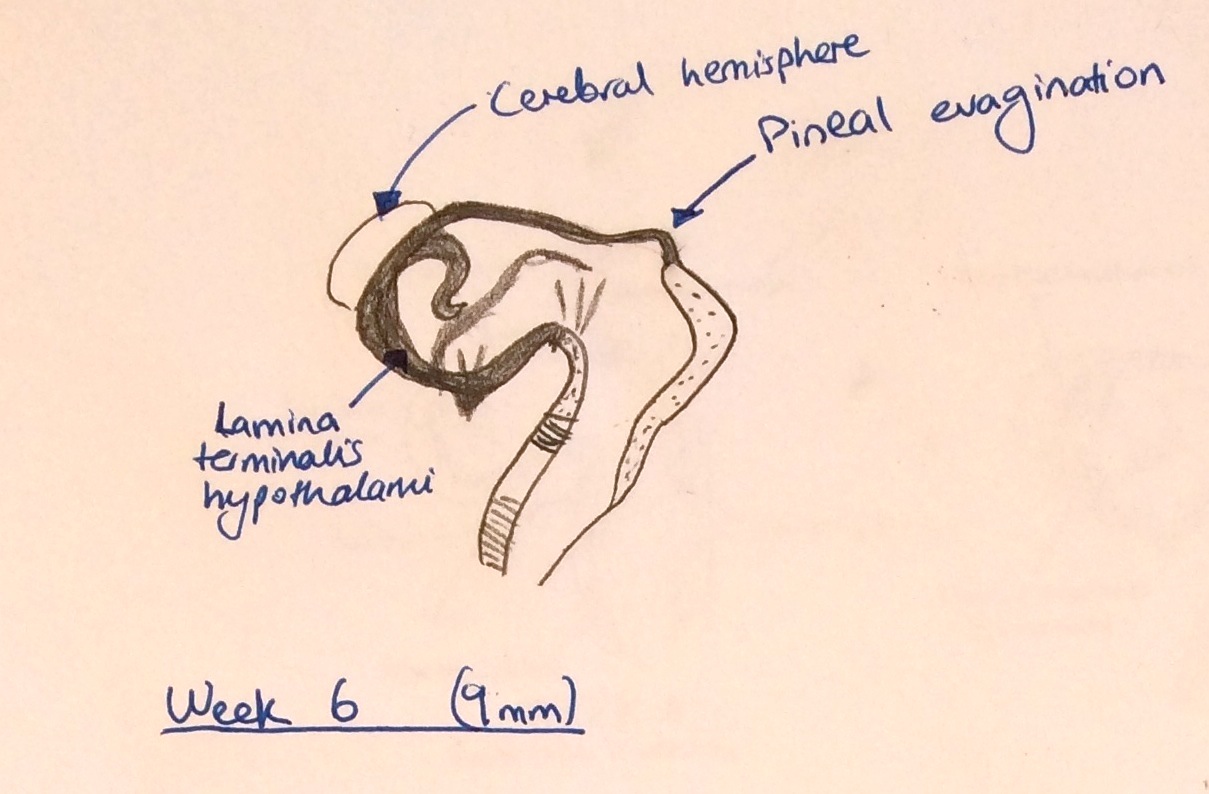
Week 6 embryonic development of CNS and emergence of pineal evagination.
This image shows the structural processes of the CNS that occur in Week 6.
This image is adapted from:[1]
References
- ↑ B. Pansky, ‘The Diencephalon, second vesicle’ Review of Medical Embryology:2011, http://discovery.lifemapsc.com/library/review-of-medical-embryology/chapter-153-the-diencephalon-second-vesicle
Copyright
Beginning six months after publication, I z3418698 grant the public the non-exclusive right to copy, distribute, or display the Work under a Creative Commons Attribution-Noncommercial-Share Alike 3.0 Unported license, as described at http://creativecommons.org/licenses/by-nc-sa/3.0/ and http://creativecommons.org/licenses/by-nc-sa/3.0/legalcode
- Note - This image was originally uploaded as part of an undergraduate science student project and may contain inaccuracies in either description or acknowledgements. Students have been advised in writing concerning the reuse of content and may accidentally have misunderstood the original terms of use. If image reuse on this non-commercial educational site infringes your existing copyright, please contact the site editor for immediate removal.
z3418698
File history
Click on a date/time to view the file as it appeared at that time.
| Date/Time | Thumbnail | Dimensions | User | Comment | |
|---|---|---|---|---|---|
| current | 13:02, 23 October 2014 | | 1,209 × 794 (165 KB) | Z3418698 (talk | contribs) | Week 6 embryonic development of CNS and emergence of pineal evagination. Adapted from <ref name=Pansky>B. Pansky, ‘The Diencephalon, second vesicle’ Review of Medical Embryology:2011, http://discovery.lifemapsc.com/library/review-of-medical-embry... |
You cannot overwrite this file.
File usage
The following page uses this file: