File:Wallin1913-fig04.jpg
From Embryology

Size of this preview: 800 × 437 pixels. Other resolution: 1,347 × 736 pixels.
Original file (1,347 × 736 pixels, file size: 122 KB, MIME type: image/jpeg)
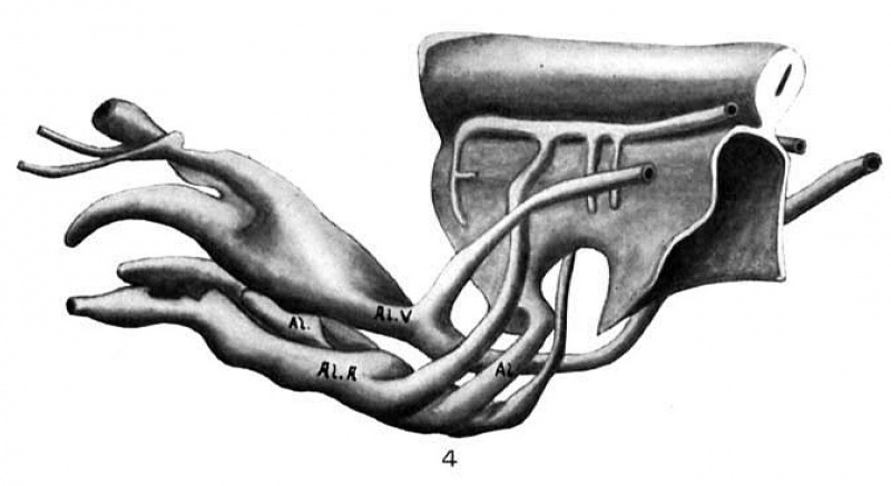
Fig. 4. Wax plate reconstruction of caudal end of the medullary tube and hind-gut with the belly stalk vessels viewed from the side
X 100.
| Historic Disclaimer - information about historic embryology pages |
|---|
 |
- Links: Fig 1 | Fig 2 | Fig 3 | Fig 4 | Fig 5 | Fig 6 | Fig 7 | Wallin 1913 | Historic Embryology Papers
Reference
Cite this page: Hill, M.A. (2024, May 11) Embryology Wallin1913-fig04.jpg. Retrieved from https://embryology.med.unsw.edu.au/embryology/index.php/File:Wallin1913-fig04.jpg
- © Dr Mark Hill 2024, UNSW Embryology ISBN: 978 0 7334 2609 4 - UNSW CRICOS Provider Code No. 00098G
File history
Click on a date/time to view the file as it appeared at that time.
| Date/Time | Thumbnail | Dimensions | User | Comment | |
|---|---|---|---|---|---|
| current | 22:05, 11 September 2015 | | 1,347 × 736 (122 KB) | Z8600021 (talk | contribs) |
You cannot overwrite this file.
File usage
The following page uses this file: