File:Uterine tube histology 05.jpg
From Embryology
Size of this preview: 600 × 600 pixels. Other resolution: 1,063 × 1,063 pixels.
Original file (1,063 × 1,063 pixels, file size: 508 KB, MIME type: image/jpeg)
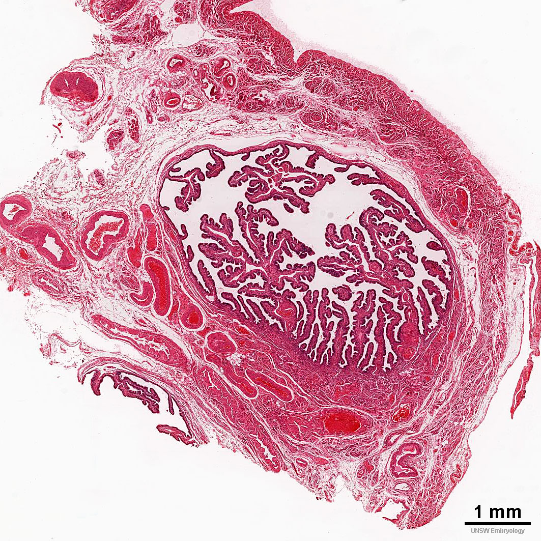
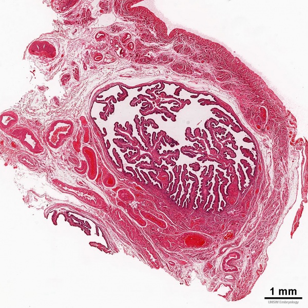
Adult Uterine Tube Histology
Uterine tube, Fallopian tube, oviduct, uterine horn.
Tube Anatomical Four Subdivisions
- Infundibulum is the funnel-shaped (up to 10 mm in diameter) end of the oviduct. Finger-like extensions of its margins, the fimbriae, are closely applied to the ovary. Ciliated cells are frequent. Their cilia beat in the direction of the ampulla of the oviduct.
- Ampulla - Mucosal folds, or plicae, and secondary folds which arise from the plicae divide the lumen of the ampulla into a very complex shape.. Fertilization usually takes place in the ampulla.
- Isthmus is the narrowest portion (2-3 mm in diameter) of the parts of the oviduct located in the peritoneal cavity. Mucosal folds are less complex and the muscularis is thick. An inner, longitudinal layer of muscle is present in the isthmus.
- Intramural part of the oviduct, which penetrates the wall of the uterus body. An inner, longitudinal layer of muscle is present.
- Links: Uterus Development
Cite this page: Hill, M.A. (2024, May 19) Embryology Uterine tube histology 05.jpg. Retrieved from https://embryology.med.unsw.edu.au/embryology/index.php/File:Uterine_tube_histology_05.jpg
- © Dr Mark Hill 2024, UNSW Embryology ISBN: 978 0 7334 2609 4 - UNSW CRICOS Provider Code No. 00098G
File history
Click on a date/time to view the file as it appeared at that time.
| Date/Time | Thumbnail | Dimensions | User | Comment | |
|---|---|---|---|---|---|
| current | 00:23, 21 April 2016 | | 1,063 × 1,063 (508 KB) | Z8600021 (talk | contribs) | |
| 00:21, 21 April 2016 | 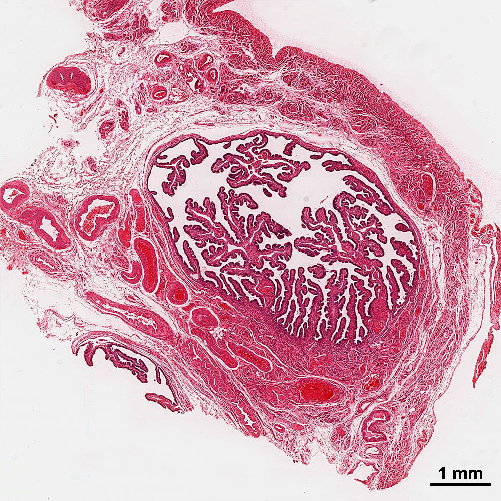 | 1,000 × 1,000 (445 KB) | Z8600021 (talk | contribs) | ==Adult Uterine Tube Histology== |
You cannot overwrite this file.
File usage
There are no pages that use this file.