File:Uterine and placental vasculature.jpg
Uterine_and_placental_vasculature.jpg (614 × 472 pixels, file size: 143 KB, MIME type: image/jpeg)
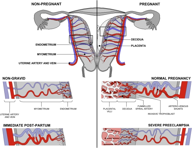
Uterine and Placental Vasculature in Non-pregnant, Pregnant and immediate Post-partum State
Diagrammatic representation of uterine and placental vasculature (red shading = arterial; blue shading = venous) in the non-pregnant, pregnant and immediate post-partum state.
Normal pregnancy is characterized by the formation of large arterio-venous shunts that persist in the immediate post-partum period.
By contrast pregnancies complicated by severe preeclampsia are characterized by minimal arterio-venous shunts, and thus narrower uterine arteries. Extravillous cytotrophoblast invasion in normal pregnancy (diamonds) extends beyond the decidua into the inner myometrium resulting in the formation of funnels at the discharging tips of the spiral arteries. Contrast with severe preeclampsia. (Prepared by Ms. Leslie Proctor, MSc.)
- Links: Figure - Uterine and placental vasculature | Uterine vascular anastomoses | Figure - Placenta spiral artery conversion | placenta
Reference
Burton GJ, Woods AW, Jauniaux E & Kingdom JC. (2009). Rheological and physiological consequences of conversion of the maternal spiral arteries for uteroplacental blood flow during human pregnancy. Placenta , 30, 473-82. PMID: 19375795 DOI.
Copyright
© 2009 Elsevier Ltd. “This is an unofficial translation of an article that appeared in an Elsevier publication. Elsevier has not endorsed this translation.”
http://www.elsevier.com/wps/find/authorsview.authors/supplementalterms1.0
Original File name: Gr2.jpg
Cite this page: Hill, M.A. (2024, April 26) Embryology Uterine and placental vasculature.jpg. Retrieved from https://embryology.med.unsw.edu.au/embryology/index.php/File:Uterine_and_placental_vasculature.jpg
- © Dr Mark Hill 2024, UNSW Embryology ISBN: 978 0 7334 2609 4 - UNSW CRICOS Provider Code No. 00098G
File history
Click on a date/time to view the file as it appeared at that time.
| Date/Time | Thumbnail | Dimensions | User | Comment | |
|---|---|---|---|---|---|
| current | 09:43, 16 August 2009 | | 614 × 472 (143 KB) | S8600021 (talk | contribs) | Diagrammatic representation of uterine and placental vasculature (red shading = arterial; blue shading = venous) in the non-pregnant, pregnant and immediate post-partum state. Normal pregnancy is characterized by the formation of large arterio-venous sh |
You cannot overwrite this file.
File usage
The following 12 pages use this file: