File:Thyroid histology 004.jpg
Original file (1,280 × 1,024 pixels, file size: 351 KB, MIME type: image/jpeg)
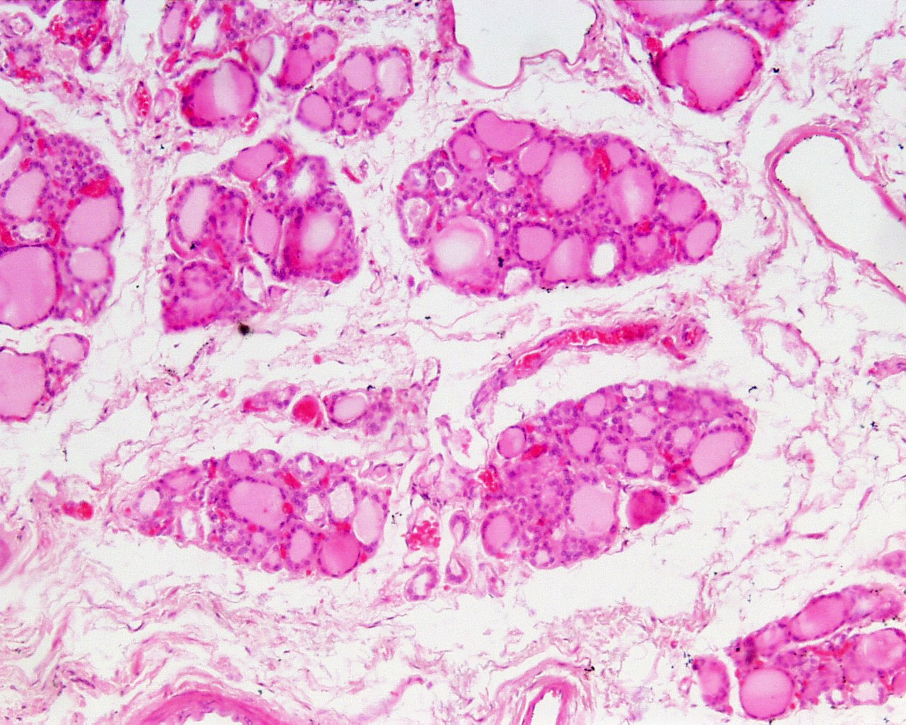
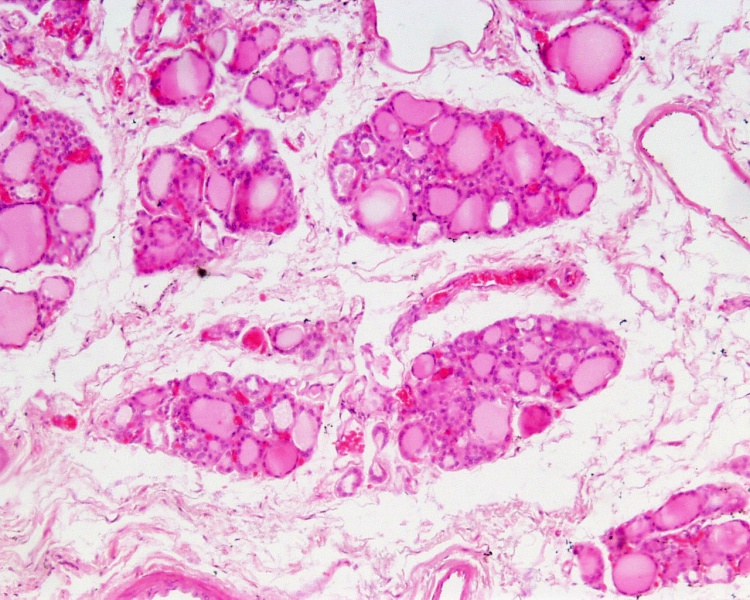
Thyroid Histology
Low power overview of the human thyroid gland histology showing follicles and connective tissue.
- consists almost entirely of rounded cysts, follicles,
- separated by small amounts interfollicular connective tissue.
- capillaries located in the interstices between the thyroid follicles.
- C cells are very difficult to identify.
- Thyroid Links: low power image | high power image | unlabeled human image | unlabeled sheep image | thyroid
Links: Histology | Histology Stains | Blue Histology images copyright Lutz Slomianka 1998-2009. The literary and artistic works on the original Blue Histology website may be reproduced, adapted, published and distributed for non-commercial purposes. See also the page Histology Stains.
Cite this page: Hill, M.A. (2024, May 21) Embryology Thyroid histology 004.jpg. Retrieved from https://embryology.med.unsw.edu.au/embryology/index.php/File:Thyroid_histology_004.jpg
- © Dr Mark Hill 2024, UNSW Embryology ISBN: 978 0 7334 2609 4 - UNSW CRICOS Provider Code No. 00098G
thy10he.jpg
File history
Click on a date/time to view the file as it appeared at that time.
| Date/Time | Thumbnail | Dimensions | User | Comment | |
|---|---|---|---|---|---|
| current | 15:49, 6 February 2013 | | 1,280 × 1,024 (351 KB) | Z8600021 (talk | contribs) | ==Thyroid Histology== Low power overview of the human thyroid gland histology showing follicles and connective tissue. * consists almost entirely of rounded cysts, follicles, * separated by small amounts interfollicular connective tissue. * capillaries |
You cannot overwrite this file.
File usage
There are no pages that use this file.