File:T2 lymphocyte EM13.jpg
Original file (781 × 795 pixels, file size: 177 KB, MIME type: image/jpeg)
T Lymphocyte Electron Micrograph
- effector T cell has relatively little rough ER but is filled with free ribosomes.
Cytotoxic T lymphocytes (CTLs) (killer T cells) - directly attack other cells carrying certain foreign or abnormal molecules on their surfaces. CTLs are especially useful for attacking viruses because viruses often hide from other parts of the immune system while they grow inside infected cells. CTLs recognize small fragments of these viruses peeking out from the cell membrane and launch an attack to kill the infected cell.
Helper T cells (Th cells) - coordinate immune responses by communicating with other cells. Some stimulate nearby B cells to produce antibodies, others call in microbe-gobbling cells called phagocytes, and still others activate other T cells.
Normal peripheral blood:
- 60 - 80% of lymphocytes are T cells
- 10 - 15% are B cells
- remainder lack both B and T cell markers (Natural killer cells, null cells).
T cell subpopulations defined by antigenic markers
- CD4+ (helper)
- CD8+ (suppressor)
- killer (cytotoxic)
- memory cells
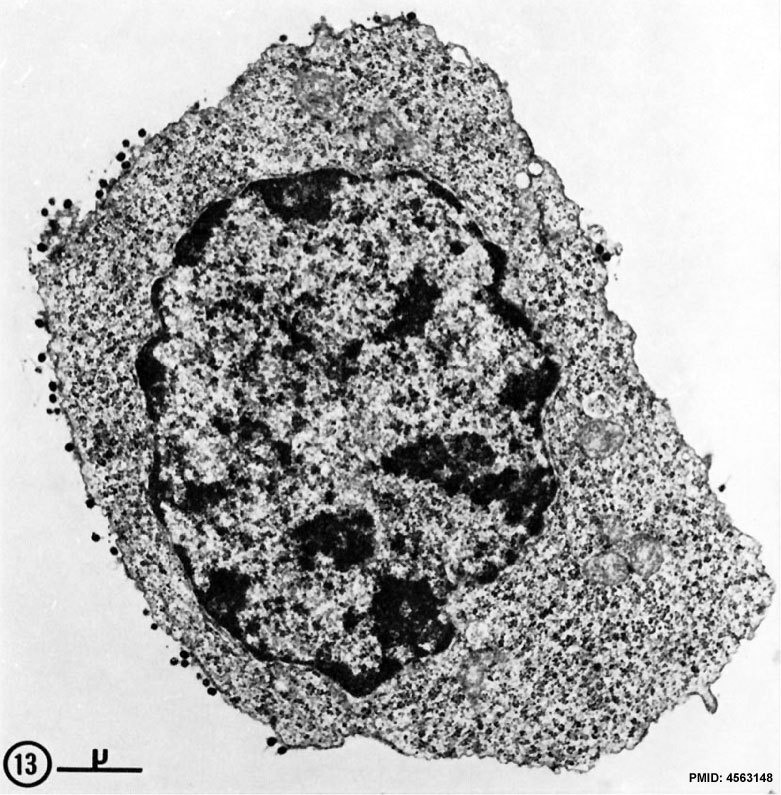
FIg. 13. Spleen cell (10 days after alloantigenic immunization with DBA/2 mastocytoma) labeled with aMSLA detected by the bridge technique with phage T4. Some of the phage heads are sectioned tangentially and therefore barely visible.
This large blast-like cell, classified as T2 lymphocyte, is mostly characterized by its very large content in polyribosomes. X 15,000. Note this T2/T3 terminology may be historic.
- Lymphocyte EM Images: T and B Lymphocytes 1 TEM | T and B Lymphocytes 2 TEM | T Lymphocyte SEM | B lymphocyte 1 TEM | B lymphocyte 2 TEM | B lymphocyte 3 TEM | Plasma Cell TEM | T2 Lymphocyte 1 TEM | T2 Lymphocyte 2 TEM | lymphocyte rosettes | T lymphocyte 1 | T lymphocyte 2 | T lymphocyte 3 | T lymphocyte 4 | T lymphocyte 5 | T lymphocyte 6 | B lymphocyte | B lymphocytes TEM | Immune System Development | Blood
Reference
Matter A, Lisowska-Bernstein B, Ryser JE, Lamelin JP & Vassalli P. (1972). Mouse thymus-independent and thymus-derived lymphoid cells. II. Ultrastructural studies. J. Exp. Med. , 136, 1008-30. PMID: 4563148
Copyright
Rockefeller University Press - Copyright Policy This article is distributed under the terms of an Attribution–Noncommercial–Share Alike–No Mirror Sites license for the first six months after the publication date (see http://www.jcb.org/misc/terms.shtml). After six months it is available under a Creative Commons License (Attribution–Noncommercial–Share Alike 4.0 Unported license, as described at https://creativecommons.org/licenses/by-nc-sa/4.0/ ). (More? Help:Copyright Tutorial)
Cite this page: Hill, M.A. (2024, April 26) Embryology T2 lymphocyte EM13.jpg. Retrieved from https://embryology.med.unsw.edu.au/embryology/index.php/File:T2_lymphocyte_EM13.jpg
- © Dr Mark Hill 2024, UNSW Embryology ISBN: 978 0 7334 2609 4 - UNSW CRICOS Provider Code No. 00098G
Some of the above text is sourced from NAIAD text
File history
Click on a date/time to view the file as it appeared at that time.
| Date/Time | Thumbnail | Dimensions | User | Comment | |
|---|---|---|---|---|---|
| current | 16:23, 22 February 2012 | | 781 × 795 (177 KB) | Z8600021 (talk | contribs) | ==T2 lymphocyte Electron Micrograph== FIg. 13. Spleen cell (10 days after alloantigenic immunization with DBA/2 mastocytoma) labeled with aMSLA detected by the bridge technique with phage T4. Some of the phage heads are sectioned tangentially and theref |
You cannot overwrite this file.
File usage
The following 8 pages use this file: