File:Streeter1921 fig11.jpg

Original file (908 × 751 pixels, file size: 103 KB, MIME type: image/jpeg)
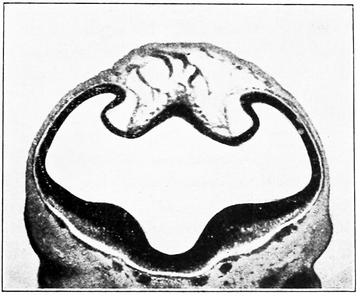
Fig. 11. Section showing the sagittal plexus in a human embryo 14 mm long
Carnegie Collection, No. 940 (slide 15 row 3. section 1)
The section shows the falciform area, the hemispheres being retracted from its lateral margins. It will be noted that there are two main plexiform vascular sheets — a superficial one near the skin and a deeper one directly against the brain-wall, the latter draining into the former by anastomosing loops. The superior sagittal sinus develops in the meshes of the superficial plexus. and the straight sinus develops in the meshes of the deep plexus over the area corresponding to the third ventricle.
- 1921 Human Brain Vascular: Fig 1 | Fig 2 | Fig 3 | Fig 4 | Fig 5 | Fig 6 | Fig 7-9 | Fig 10 | Fig 11 | Fig 12 |Fig 13 | Fig 14 | Fig 15 | Fig 16 | Fig 17 | Fig 18 | Fig 19 | Fig 20 | Fig 21 | Fig 22 | Fig 23 | Fig 24 | Fig 25 | Fig 26 | Fig 27 | Plate 1 - embryos 4 mm to birth | Plate 2 - embryo 4 mm | Plate 3 - embryo 11.5 mm | Plate 4 - embryo 21 mm | Plate 5 - embryo 43 mm | Carnegie No.24 | George Streeter
| Historic Disclaimer - information about historic embryology pages |
|---|
 |
Reference
Streeter GL. The developmental alterations in the vascular system of the brain of the human embryo. (1921) Contrib. Embryol., Carnegie Inst. Wash. 8:7-38.
Cite this page: Hill, M.A. (2024, April 27) Embryology Streeter1921 fig11.jpg. Retrieved from https://embryology.med.unsw.edu.au/embryology/index.php/File:Streeter1921_fig11.jpg
- © Dr Mark Hill 2024, UNSW Embryology ISBN: 978 0 7334 2609 4 - UNSW CRICOS Provider Code No. 00098G
File history
Click on a date/time to view the file as it appeared at that time.
| Date/Time | Thumbnail | Dimensions | User | Comment | |
|---|---|---|---|---|---|
| current | 11:25, 19 January 2017 | | 908 × 751 (103 KB) | Z8600021 (talk | contribs) | |
| 03:33, 21 April 2012 | 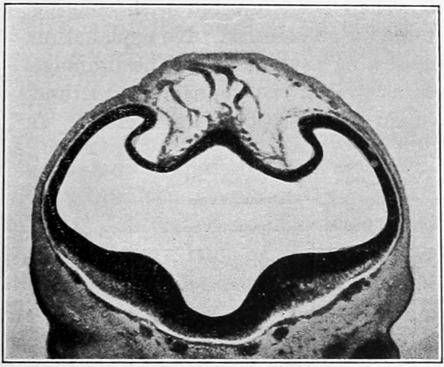 | 908 × 751 (124 KB) | Z8600021 (talk | contribs) | Fig. 11. Section showing the sagittal plexus in a human embryo 14 mm. long (Carnegie Collection, No. slide l.j. rr.w .3. section 1). The section shows the falciform area, the hemi.spheres beiiit; riu.n u-il from its lateral margins. It will be noted that |
You cannot overwrite this file.
File usage
The following page uses this file: