File:Streeter1920 01.jpg
Original file (723 × 1,000 pixels, file size: 63 KB, MIME type: image/jpeg)
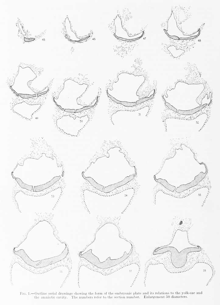
Fig. 1. Outline serial drawings showing the form of the embryonic plate and its relations to the yolk-sac and the amniotic cavity
Outline serial drawings showing the form of the embryonic plate and its relations to the yolk-sac and the amniotic cavity. The numbers refer to the section number. Enlargement 50 diameters.
Sections 44 and 45 pass tangentially through the extreme rostral portion of the anmion. In section 45 the amniotic cavity makes its first appearance. Dorsally it is loosely adherent to the chorionic membrane.
Section 46 passes through the rostral end of the amniotic cavity and shows the obliquely cut rostral margin of the embryonic plate. An irregular strand of mesodermal cells can be seen everywhere investing the amniotic membrane and embryonic shield.
Sections 47 to 49 penetrate more deeply into the amniotic cavity showing the folds of the amniotic membrane. Ventral to the amniotic cavity the yolk-sac makes its appearance.
Section 50: A more detailed drawing of this section is shown in figure 16, plate 4. The amniotic cavity is still larger than the yolk cavity. The anmiotic membrane is folded so that it can be seen in both transverse and tangential sections. Where cut transversely it appears as a single layer of flattened ectodermal cells closely invested by an irregular layer of mesoderm. Dorsally, loose strands of mesoderm extend toward the chorionic membrane. Around its lateral margin the amniotic membrane bends sharply to become the embryonic plate. In its more lateral portions the embryonic plate consists of one or two layers of cylindrical ectodermal cells. The nuclei for the most part are toward the bases of the cells. More centrally the number of layers is increased to 3 or 4. Owing to the tangential direction of the sections, the middle portion of the embryonic plate appears thicker than it actually is. Ventral to the embryonic plate, strands of mesoderm extend between it and the yolk-sac. The mesoderm seems to be more adherent to the ectoderm of the embryonic plate than to the entoderm of the yolk-sac; between it and the latter there is a series of roomy clefts. The wall of the yolk-sac consists of a rather poorly defined strand of protoplasm with large, round, and oval nuclei, arranged irregularly in two layers. Some of these stam intensely, others are pale. Cell-boundaries can not be well made out. Within the yolk-sac there is a considerable amount of finely granular coagulum similar to that in the exoccelom. The amniotic cavity is perfectly clear. Apparently the outer row of nuclei represents the investment of mesoderm.
Sections 51 and 52: The space between the amnion and chorionic membrane is bridged by a mass of mesoderm more dense than in the previous sections, so that the sections are now definitely in the region of the body-stalk. Among these cells may occasionally be seen a group arranged in circular formation, so as to form a disconnected endothelial-like space. These spaces are for the most part empty, but now and then they inclose one or more cells. The amniotic membrane is much the same as in the previous sections. The embrjonic plate is cut obliquely. The central part is uniform in appearance, showing no evidence of an neurenteric canal. The margin towards the amniotic cavity is covered on each side by a lateral sulcus which demarcates a transitional portion intervening between the embryonic plate and the amniotic membrane. This portion resembles the rhombic lip, to which is attached the tela choroidea in the hind-brain of the adult. About one-third of the distance between this lateral sulcus and the middle line is another groove, less marked, but which seems to be fairly constant throughout the successive sections. In the middle line there is no groove. Between the two sulci on each side the embryonic plate bulges into the lumen of the amniotic cavity, resulting in a longitudinal ridge which can be traced backward to about the region of the primitive groove. In the embedding of the specimen the tissue became brittle and an occasional crack is found crossing the embryonic plate. The mesodenn ventral to the embryonic plate shows pointy of intimate attachment to the latter, particularly in the lateral portions of the plate. Strands of mesoderm cross from the embryonic plate to the yolk-sac, forming trabeculae, between which is a series of clear, round spaces. Laterally, these spaces are continuous with the cleft that intervenes between the mesoderm and the wall of the yolk-sac, extending about one-quarter of the distance toward the ventral pole. The two layers of the wall of the yolk-sac, the endoderm and mesoderm, are more distinct than in the previous sections. No indication of blood islands is seen in this region. The content of the yolk-sac resembles the granular magma seen in the exoccelom and is perhaps slightly greater in amount.
Section 53 shows very well the attachment between the amnion and the chorionic membrane. The mesodermal cells are closely clustered around the anmion. The apex of the amnion is cut tangentially and so stands out in marked contrast to the mesoderm. The transition from the amnion to the embryonic plate is clearly shown on the left side of the section, the transitional portion being made up mostly of one layer of entodermal cells. The embryonic plate is everywhere clearly separated from the yolk-sac by the intervening mesoderm, which at several points seems adherent to it.
Section 54: In this section the amnion comes in contact with the chorionic membrane. The amniotic ectoderm does not show any connection with the chorionic epithelium.
Section 55: In the body-stalk there is seen an endothelial-like space within the lumen of which a cluster of 7 nuclei projects. The mesoderm lie between the embryonic plate and the yolk-sac is more closely attached to the former than to the latter.
Section 56: (Compare fig. 16, plate 4.) This section is particularly good for showing the relations between the amniotic membrane and the mesoderm. The former is nearly everywhere cvit in transverse section except at its extreme tip. The mesoderm is arranged as a membrane, closely investing the amnion and extending a short distance on the body-stalk. Ventrally it extends downward to inclose the yolk-sac, where it can be traced as a separate lamina for about one-half the distance to the ventral pole. Lying free in the exocoelomic space, at the; junction of the amnion with the body-stalk, is a small, empty, endothelial cavity. This can be traced only through two sections. It is surrounded solely by finely granular coagulum. The mesoderm between the embryonic plate and the yolk-sac is adherent at many points to both. The wall of the yolk-sac is cut obliquely for the most part. Its cavity now appears somewhat larger than the amniotic cavity. No blood islands are seen.
Section 57: A very intimate relation exists between the lateral wings of the embryonic plate and the subjacent mesoderm. In the body-stalk, near the tip of the amnion, there is a small mass of cells wliich ajiparently are ectodermal and may represent a bud from the amniotic ectoderm, which ap])ears detacheil on account of the tangential ilirection of the sections.
Section 58: According to the memoranda obtained from Dr. Willier, two sections through the embryo were lost . On account of the abrupt transition between sections 57 and 58, it would seem probable that the sections are missing at this point. The abruptness is due partly also to the curve in the longitudinal axis of the embryonic plate, so that the plate is cut in this and the succeeding two sections in a markedly tangential direction. In this section the body-stalk is more condensed than heretofore and is fairly well inclosed by a membranous arrangement of the inesotlemi. It contains in its center the tip of the allantoic duct. At one point there is a slight indication of a lumen. The anmiotic cavity has becomi; considerably contracted and conforms in a blunt manner to the fonn of the body-stalk. An intermediate plate still exists between the amniotic membrane and the embryonic plate. This is the first section in which a sharp groove appears in the median line of the embryonic plate — the primitive groove. At this point the ectoderm, mesoderm, and endoderm of the yolk-sac form one continuous mass, which corresponds to the primitive node of Hensen. The extent of this area is exaggerated, owing to the obliqueness of the section. In the ventral part of the yolk-sac a cluster of cells, apparently representing bloodislands, can be recognized.
| Historic Disclaimer - information about historic embryology pages |
|---|
 |
- Paper Links: Fig 1 | Fig 2 | Fig 3 | Fig 4 | Fig 5 | Fig 6 | Fig 7 | Fig 8 | Fig 9 | Fig 10 | Fig 11 | Fig 12 | Fig 13 | Fig 15 | Fig 16 | Table 1 | Chart 1 | Chart 2 | Chart 3 | Plate 1 | Plate 2 | Plate 3 | Plate 4 | Plate 5 | Plate 6 | Plate 7 | Paper | Contributions to Embryology
Reference
Streeter GL. A human embryo (Mateer) of the pre-somite period. (1920) Contrib. Embryol., Carnegie Inst. Wash. Publ. 272, 9: 389-424.
Cite this page: Hill, M.A. (2024, April 27) Embryology Streeter1920 01.jpg. Retrieved from https://embryology.med.unsw.edu.au/embryology/index.php/File:Streeter1920_01.jpg
- © Dr Mark Hill 2024, UNSW Embryology ISBN: 978 0 7334 2609 4 - UNSW CRICOS Provider Code No. 00098G
File history
Click on a date/time to view the file as it appeared at that time.
| Date/Time | Thumbnail | Dimensions | User | Comment | |
|---|---|---|---|---|---|
| current | 09:35, 7 April 2012 | | 723 × 1,000 (63 KB) | Z8600021 (talk | contribs) | :Links: ===Reference=== Streeter G.L. A Human Embryo (Mateer) Of The Presomite Period. Contributions to Embryology Carnegie Institution No.43 (1920) pp389-424, 4 text-figures and 7 plates. {{Historic Disclaimer}} Category:George Streeter |
You cannot overwrite this file.
File usage
The following page uses this file: