File:Streeter1906 fig03.jpg

Original file (1,472 × 1,943 pixels, file size: 285 KB, MIME type: image/jpeg)
Fig. 3. Development of a semicircular canal
A, B, C, and D represent transverse sections of the superior canal (x240), taken at corresponding points from embryos No. 163, 9 mm.; No.109, 11 mm.; No. 229, 21 mm., and No. 86, 30 mm.
Aa, Ab, Ba,and Bb are explanatory drawings of lower magnification to show the ear vesicle and the situation and shape of sections from which A and B are taken.
| Historic Disclaimer - information about historic embryology pages |
|---|
 |
- Mall 1906 Links: Fig 1. 14mm Embryo | Fig 2. 30mm Embryo | Fig 3. Semicircular canal | Fig 4. Membranous Labyrinth | Fig 5. Acoustic nerve complex | Fig 6. Facial-acoustic Complex | Fig 7. Facial Nerve Pig Embryo 20 cm | Fig 8. Geniculate Ganglion | Plate 1. Human Embryo 4 to 20 mm | Plate 2. Human Embryo 30 mm | Membranous Labyrinth and Nerves

Fig 1 Membranous Labyrinth Human Embryo 14 mm
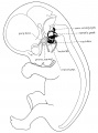
Fig 2 30mm Embryo
Fig 3 Semicircular canal
Fig 4 Membranous Labyrinth Growth
Fig 5 Acoustic nerve complex

Fig 6 Facial-acoustic Complex Human Embryo 7 mm
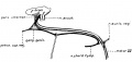
Fig 7 Facial Nerve Pig Embryo 20 cm
Fig 8 Geniculate Ganglion Human Embryo 30 mm
Plate 1. Membranous Labyrinth Human Embryo 4 to 20 mm
Plate 2. Membranous Labyrinth Human Embryo 30 mm


Reference
Streeter GL. On the development of the membranous labyrinth and the acoustic and facial nerves in the human embryo. (1906) Amer. J Anat. 6:139-165.
Cite this page: Hill, M.A. (2024, May 15) Embryology Streeter1906 fig03.jpg. Retrieved from https://embryology.med.unsw.edu.au/embryology/index.php/File:Streeter1906_fig03.jpg
- © Dr Mark Hill 2024, UNSW Embryology ISBN: 978 0 7334 2609 4 - UNSW CRICOS Provider Code No. 00098G
File history
Click on a date/time to view the file as it appeared at that time.
| Date/Time | Thumbnail | Dimensions | User | Comment | |
|---|---|---|---|---|---|
| current | 17:10, 23 July 2015 | | 1,472 × 1,943 (285 KB) | Z8600021 (talk | contribs) | |
| 17:10, 23 July 2015 | 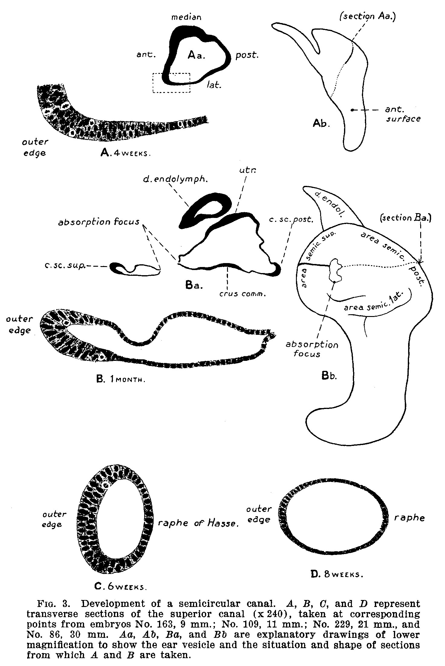 | 1,472 × 2,200 (384 KB) | Z8600021 (talk | contribs) |
You cannot overwrite this file.
File usage
The following 14 pages use this file:
- Carnegie stage 23
- Hearing - Inner Ear Development
- Paper - On the development of the membranous labyrinth and the acoustic and facial nerves in the human embryo
- File:Streeter1906 fig01.jpg
- File:Streeter1906 fig02.jpg
- File:Streeter1906 fig03.jpg
- File:Streeter1906 fig04.jpg
- File:Streeter1906 fig05.jpg
- File:Streeter1906 fig06.jpg
- File:Streeter1906 fig07.jpg
- File:Streeter1906 fig08.jpg
- File:Streeter1906 plate01.jpg
- File:Streeter1906 plate02.jpg
- Template:Streeter1906 figures