File:Streeter030.jpg
Original file (774 × 1,000 pixels, file size: 78 KB, MIME type: image/jpeg)
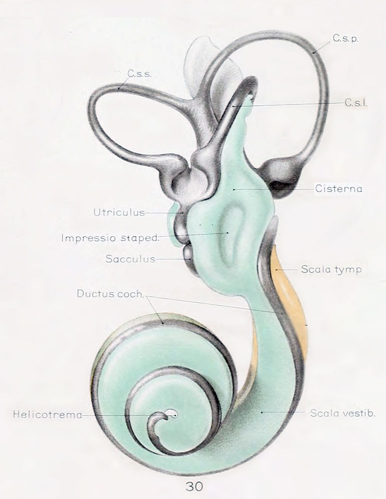
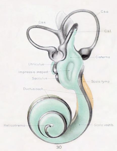
Fig. 30. Lateral view of left membranous labyrinth and the periotic spaces in a human fetus 130 mm CRL
Lateral view of a wax-plate reconstruction of the left membranous labyrinth and the periotic spaces in a human fetus 130 mm. crown-rump length (Carnegie Collection, No. 1018), enlarged 11.4 diameters.
The cistern and scala vestibuli are shown in green and the scala tympani is shown in orange, as in the previous figures.
The cartilaginous stapes was removed from this model and the oval impression that it makes on the cistern can be plaiidy seen. The cistern has spread over the top of the utricle and part way along the lateral semicircular duct. The scala vestibuli extends to the tip of the cochlear duct, where it communicates with the scala tympani, thus forming the helicotrema.
The figures shown on this plate 4 represent a series of median and lateral views of wax-plate reconstructions of the membranous labyrinth and the surrounding periotic tissue-spaces. They illustrate under the same scale of enlargement three typical stages in the development of these spaces.
Abbreviations
- C. s. 1. - ductus semicircuiaris lateralis
- C. s. p. - ductus semicircularis posterior
- C. s. s. - ductus semicircularis superior
- Duct, coch. - ductus cochlearis
- Impressio rotund. - area opposite the fenestra cochleae
- Impressio staped., area in contact with base of stapes
- Saccus endol. - saccus endolymphaticus
- Scala tymp. - scala tynipani
- Scala vestib. - scala vestibule.
Reference
Streeter G.L. The histogenesis and growth of the otic capsule and its contained periotic tissue-spaces in the human embryo Contributions to Embryology Carnegie Institution No.20 (1918) pp5-54, 4 text-figures and 4 plates.
File history
Click on a date/time to view the file as it appeared at that time.
| Date/Time | Thumbnail | Dimensions | User | Comment | |
|---|---|---|---|---|---|
| current | 21:52, 22 April 2012 | | 774 × 1,000 (78 KB) | Z8600021 (talk | contribs) | ====Fig. 30==== Lateral view of a wax-plate reconstruction of the left membnmoiis labyrinth and the periotic spaces in a human fetus 130 mm. crown-rump length (Carnegie Collection, No. 1018), enlarged 11.4 diameters. The cistern and scala vestibuli are sh |
You cannot overwrite this file.
File usage
The following 2 pages use this file: