File:Spermatozoa tail cross-section cartoon.jpg
Spermatozoa_tail_cross-section_cartoon.jpg (429 × 429 pixels, file size: 43 KB, MIME type: image/jpeg)
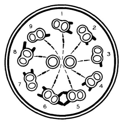
A schematic representation of a typical sperm tail in cross-section
This image is from an historic paper that identified features of the spermatozoa microtubule organisation by EM.
- The outlines of the two central filaments are circular, those of the nine peripheral filaments are of a more complex morphology.
- There are projections between the filaments: the "spokes" in radial direction, here represented by interrupted lines, and the "arms" of the peripheral filaments drawn as solid lines. It is to be noted that the two subunits of one peripheral filament differ.
- The subunit in the clockwise direction is characterized by its slightly larger radial diameter, the connection with the arms as well as with the spokes, and by the tilting of the peripheral filaments that gives the clock- wise side a slightly more central position than the counter-clockwise side.
- The asymmetry of this fila- ment arrangement has made it possible to assign an index number to each of the peripheral filaments. This was done according to the following system.
- Filament 1 is the filament that is located at an equal distance from the centers of the two inner filaments, and the increasing numbers are given to the filaments in the direction of the arms.
- The arms from filament 5 are met by projections in a counter-clockwise direction from filament 6, and the four units seem to form a complex bridge. Further details are presented in the text.
(Drawing by John Spurbeck.)
References
<pubmed>13654448</pubmed>| PDF
Copyright
Rockefeller University Press - Copyright Policy This article is distributed under the terms of an Attribution–Noncommercial–Share Alike–No Mirror Sites license for the first six months after the publication date (see http://www.jcb.org/misc/terms.shtml). After six months it is available under a Creative Commons License (Attribution–Noncommercial–Share Alike 4.0 Unported license, as described at https://creativecommons.org/licenses/by-nc-sa/4.0/ ). (More? Help:Copyright Tutorial)
Text Fig.1 PMID 13654448
File history
Click on a date/time to view the file as it appeared at that time.
| Date/Time | Thumbnail | Dimensions | User | Comment | |
|---|---|---|---|---|---|
| current | 06:09, 16 March 2012 | | 429 × 429 (43 KB) | Z8600021 (talk | contribs) | ==A schematic representation of a typical sperm tail in cross-section== This image is from an historic paper that identified features of the spermatozoa microtubule organisation by EM. * The outlines of the two central filaments are circular, those of |
You cannot overwrite this file.
File usage
There are no pages that use this file.