File:Smith1961 fig01-13.jpg
Original file (1,717 × 2,594 pixels, file size: 565 KB, MIME type: image/jpeg)
Figure 2
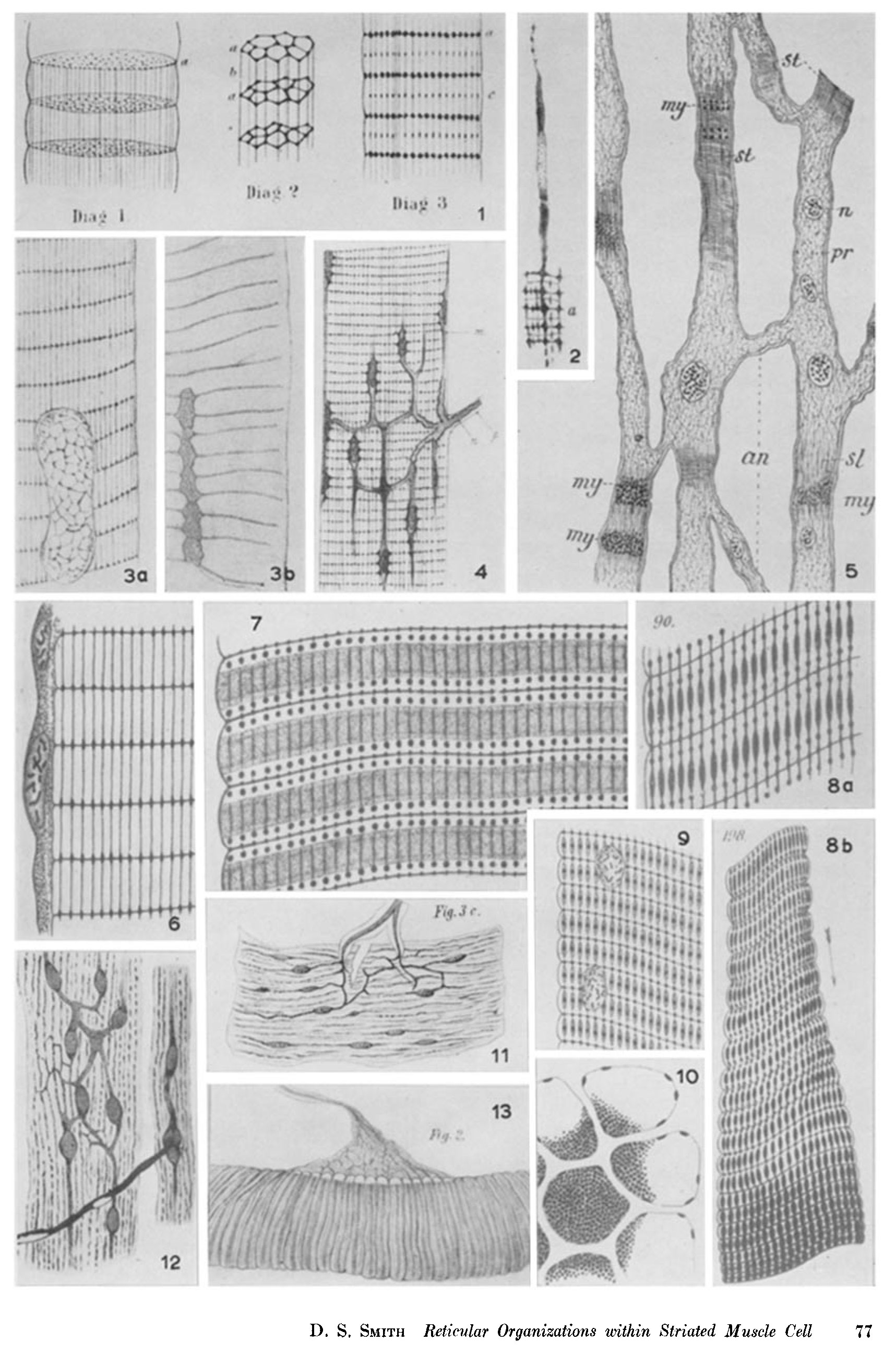
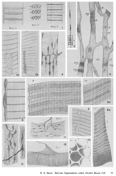
A figure from the work of Brerner (l882) in which he developed the hypothesis that the muscle Eber is composed of separate «corpuscles" (actually the scattered nuclei), each of which was thought to be produced into a protoplasmic reticulum of «Inuscle rods." This diagram represents a single corpuscle with a portion of its network, which was visualized as bearing alternately thick and thin transverse Elaments (the Z and M bands of modern terminology), linked by longitudinal connectives
Figures 3 a, 3 b, and 4
Marshall (1888) shared the view of Thanhoifen Gerlach, and others that the nerves reaching the Eber connect with the Elarnents of the postulated reticulum, which were visualized as stemming from discrete «muscle corpusles« (Fig. 4). These corpuscles in fact represent the Eber nuclei, and the supposed derivation of transverse trabeculae from them is shown in Fig. 3 ö- (Marsha1l, l890), while in Fig. 3 c: the longitudinal connecting elements are included; the striation of the Eber was thus attributed to the precise geometrical alignment of these two arrays.
Figure 5
A diagTam from Carnoy (l884), illustrating his concept of muscle structure: that the muscle cell differs from others primarily in the degree of order shown by its protoplasmic «reticulum." In this case, Carnoy imagined that he had observed the transformation of cells of the gut of Hydropfkifur into muscle, and the bands occurring where the «reticulum« became oriented into a grid of crossmeshes (m)-) were thought to represent regions where the «1nyosin" was concentrated in the «enchylema«’ and was deposited in the form of granules. The «reticular hypothesis" of protoplasmic organization put forward by Carnoy was supported and elaborated by Van Gehuchten and his followers in their work on the structure of striated muscle.
Figures 6 sro 10
Van Gehuchten, following Carnoy, was the chief proponent of the "fiuid matrix hypothesis" of muscle structure. He believed that the Ebrils were the coagulation products of a «myosin-rich" fluid which, in the living Eber, surrounded a reticular array of more solid elements, considered to be the contractile portion. His views are illustrated in the Egures here reproduced from his Erst memoir (l886). His ideas initially received much attention, and Egures from the work of several of his contemporaries who supported his views are shown elsewhere on this plate.
Fig. S. Part of a Eber from the crayEsh Arke-Jus showing the precisely organized reticulum thought to remain after extraction of the fluid matix
Fig. 7. Illustrates Van Gehuchtecks concept of the appearance of a living Eber from the beetle Mefoimxxfsrk The transverse reticulum (Z band) is evident, also the longitudinal connectives in the A band region, the optical properties and density of which were attributed to localized concentration of the matrix. The rows of dots in the clear region (I band), which may represent sarcosomes, were termed «accessory discs" by Van Gehuchten and were believed to be local thickenings in the longitudinal trabeculae, which disappeared on contraction (see Fig. 8 L7).
Fig. 8 a. A Eber corresponding to the last, after Exatiotr The coagulated fluid matrix was supposedly deposited on the meshes of the reticulum in the «dark band« (A band), where the Elaments were therefore thicker.
Fig. 8 III. A Eber from the larva of Melolontkcz showing a «Exed contraction wave." This Egure illustrates Van Gehuchtecks concept of contraction as a shortening and thickening of the longitudinal links of the reticulum, resulting in the appearance of dark contraction bands
Fig. 9. Illustrates a portion of a Eber of Not— Lade: after alcohol Exation Van Gehuchten described numerous variations in structural detail of the reticulum; in this instance the M band is represented by a dense dot at the center of each longitudinal trabecula (compare with Fig. 8 e).
Fig. l0. Representing a transverse Section through a group of alcoholdixed Ebers of Meile— Lenz-Um muscle, showing their comp0nent myoEbrils. Van Gehuchten admitted the existence of such Ebrils in the living Eber only in the case of «dissociable" flight rnuscles of certain insects.
Figure 11. An illustration from Thanhoffer (1882) of a nerve ending in gold impregnated frog muscle. He believed that in this tissue, the nerve was subdivided into longitudinal filaments which connected with nuclei lying beneath the sarcolemma rather than with an internal reticulum as was believed to be the case in insect muscle.
Figure 12. Diagrarn of gold impregnated frog muscle from Gerlach (1877), who believed, with Marsha1l, Bremer, and others, that the fiber is composed of separate bodies or cells associated with a reticulum, which connect directly (as in this iigure) with nerve branches at the end plate.
Figure 13. Here, in a fiber from the beetle Hydropfeifar, the end plate was described by ThanhoHer as containing a network, the elements of which apparently passed into Krauseks lines (Z bands).
The reticula described and illustrated in this plate do not correspond in any way to the definitive sarcoplasmic reticulum of Fusari, Cajal, Veratti, and others, but were the product of a basic misconception of muscle structure, in which the striations of the fibrils and the interstices between them were mistaken for transverse and longitudinal Hlaments, embedded in a homogeneous matrix.
Figure 14. MacCallum (1897) described the sareoplasm of human and other heart muscle as being divided into a series of (:haInbers deHned by radial and transverse membranes, arranged around the ftbrils in a rosette pattern as in the diagram reproduced here. Veratti considered it possible that what MacCallum believed to be Sections through the membranes of sarcoplasmic discs or chambers were in reality Hlamentous elements of the reticulum, though from the fixation MacCallum employed this seems unlikely
Copyright
Rockefeller University Press - Copyright Policy This article is distributed under the terms of an Attribution–Noncommercial–Share Alike–No Mirror Sites license for the first six months after the publication date (see http://www.jcb.org/misc/terms.shtml). After six months it is available under a Creative Commons License (Attribution–Noncommercial–Share Alike 4.0 Unported license, as described at https://creativecommons.org/licenses/by-nc-sa/4.0/ ). (More? Help:Copyright Tutorial)
Cite this page: Hill, M.A. (2024, April 30) Embryology Smith1961 fig01-13.jpg. Retrieved from https://embryology.med.unsw.edu.au/embryology/index.php/File:Smith1961_fig01-13.jpg
- © Dr Mark Hill 2024, UNSW Embryology ISBN: 978 0 7334 2609 4 - UNSW CRICOS Provider Code No. 00098G
File history
Click on a date/time to view the file as it appeared at that time.
| Date/Time | Thumbnail | Dimensions | User | Comment | |
|---|---|---|---|---|---|
| current | 09:07, 2 September 2018 | | 1,717 × 2,594 (565 KB) | Z8600021 (talk | contribs) |
You cannot overwrite this file.
File usage
There are no pages that use this file.