File:Shaner1945 fig02.jpg
Original file (1,280 × 1,026 pixels, file size: 155 KB, MIME type: image/jpeg)
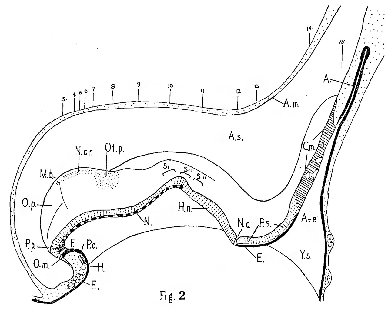
Fig. 2. Sagittal graphic reconstruction
X 60.
Key to abbreviations used in Figs. 2 to 20.
Nos. 3 to 15, position of sections shown in Figs. 3 to 15; A., allantois; A.e., allantoenteric diverticulum, Arn., amnion; A.s., amniotic sac; C.m., cloacal membrane; E., entoderm; F., fore-gut; H., heart; H.n., Henson’s node; m.b., midbrain bend; N., notochord; N.c., nenrenteric canal; N.cr., neural crest; O.m., oral membrane; O.p., optic primordium; 02!. p., otic placoole; P.c., pericardial cavity; P.p., prechordal plate; P.s., primitive streak; Y.s., yolk sac; S.I., II., III.,. somites; T.s., terminal sinus.
Reference
Shaner RF. A human embryo of two to three pairs of somites. (1945) Canad. J. Res. 23: 235-243.
Cite this page: Hill, M.A. (2024, May 2) Embryology Shaner1945 fig02.jpg. Retrieved from https://embryology.med.unsw.edu.au/embryology/index.php/File:Shaner1945_fig02.jpg
- © Dr Mark Hill 2024, UNSW Embryology ISBN: 978 0 7334 2609 4 - UNSW CRICOS Provider Code No. 00098G
File history
Click on a date/time to view the file as it appeared at that time.
| Date/Time | Thumbnail | Dimensions | User | Comment | |
|---|---|---|---|---|---|
| current | 13:01, 30 July 2017 | | 1,280 × 1,026 (155 KB) | Z8600021 (talk | contribs) | |
| 13:00, 30 July 2017 | 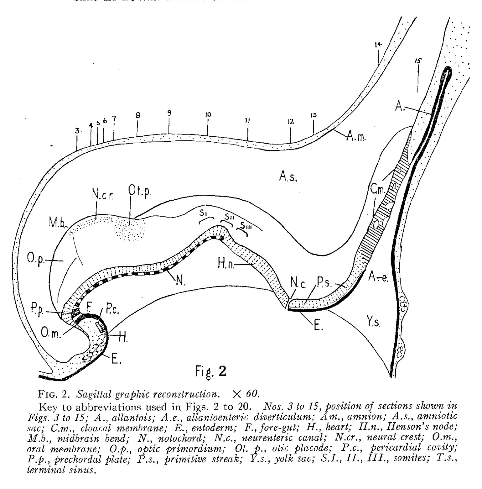 | 1,539 × 1,560 (318 KB) | Z8600021 (talk | contribs) |
You cannot overwrite this file.
File usage
The following 2 pages use this file: