File:Sabin1909 fig13.jpg
Sabin1909_fig13.jpg (640 × 547 pixels, file size: 114 KB, MIME type: image/jpeg)
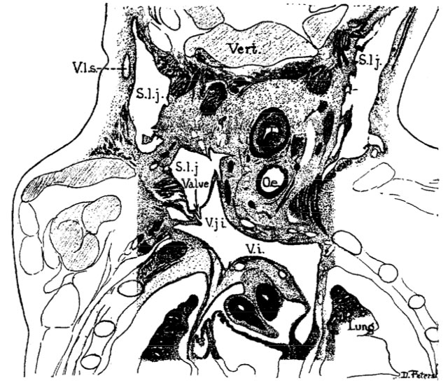
Fig. 13. Human embryo measuring 36 mm
Fig. 13. Coronal section through the jugular lymph sacs in a human embryo of 36 mm.
Mall collection, No. 86. x about 11.
The level of the section is shown on the reconstruction of Fig. 21.
The section shows the complete lymph sac on the right side and is cut to show the valve on the left S. l. j., saccus lymphaticus jugularis ; V. i., V. innominata ; V. j. F, V. jugularis interna;‘V. 1. s., vasa lymphatica superficialis.
Reference
Florence R. Sabin, The lymphatic system in human embryos, with a consideration of the morphology of the system as a whole. American Journal of Anatomy Volume 9, Issue 1, pages 43–91, 1909
File history
Click on a date/time to view the file as it appeared at that time.
| Date/Time | Thumbnail | Dimensions | User | Comment | |
|---|---|---|---|---|---|
| current | 14:16, 30 March 2011 | | 640 × 547 (114 KB) | S8600021 (talk | contribs) | ==Fig 4. Human embryo measuring 10.5 mm== Mall collection, No. 109 '''Jugular Sacs''' * The relation of the sac to the venous system as a whole is shown, which is a just external to the internal jugular vein reconstruction from serial sections. * The |
You cannot overwrite this file.
File usage
There are no pages that use this file.