File:Rugh 157.jpg
Original file (993 × 1,000 pixels, file size: 183 KB, MIME type: image/jpeg)
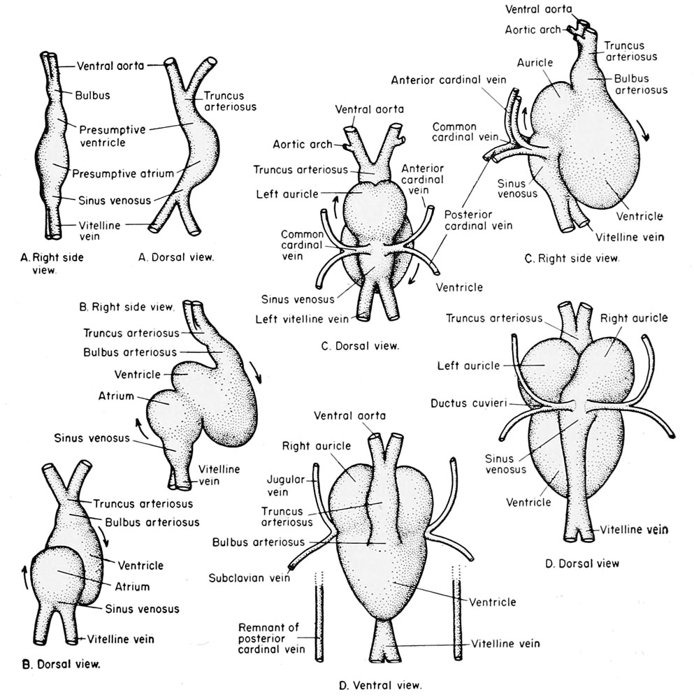
Development of the amphibian heart
The early differentiated heart consists of a simple tube which becomes divided into three chambers. The fused pair of vitelline veins becomes the meatus venosus which empties into the first division of the heart, the sinus venosus. Then follows (more anteriorly) the thinwalled atrium, the thick-walled ventricle, and finally the bulbus arteriosus (bulbus aortae) which leads to the ventral aorta or truncus arteriosus.
- Links: The Frog Heart
| Historic Disclaimer - information about historic embryology pages |
|---|
 |
Reference
Rugh R. Book - The Frog Its Reproduction and Development. (1951) The Blakiston Company.
Cite this page: Hill, M.A. (2024, May 11) Embryology Rugh 157.jpg. Retrieved from https://embryology.med.unsw.edu.au/embryology/index.php/File:Rugh_157.jpg
- © Dr Mark Hill 2024, UNSW Embryology ISBN: 978 0 7334 2609 4 - UNSW CRICOS Provider Code No. 00098G
File history
Click on a date/time to view the file as it appeared at that time.
| Date/Time | Thumbnail | Dimensions | User | Comment | |
|---|---|---|---|---|---|
| current | 10:40, 26 April 2013 | | 993 × 1,000 (183 KB) | Z8600021 (talk | contribs) | {{Rugh1951 footer}} Category:Heart Category:Cardiovascular |
You cannot overwrite this file.
File usage
The following 2 pages use this file: