File:Rugh 007.jpg
Original file (965 × 1,200 pixels, file size: 220 KB, MIME type: image/jpeg)
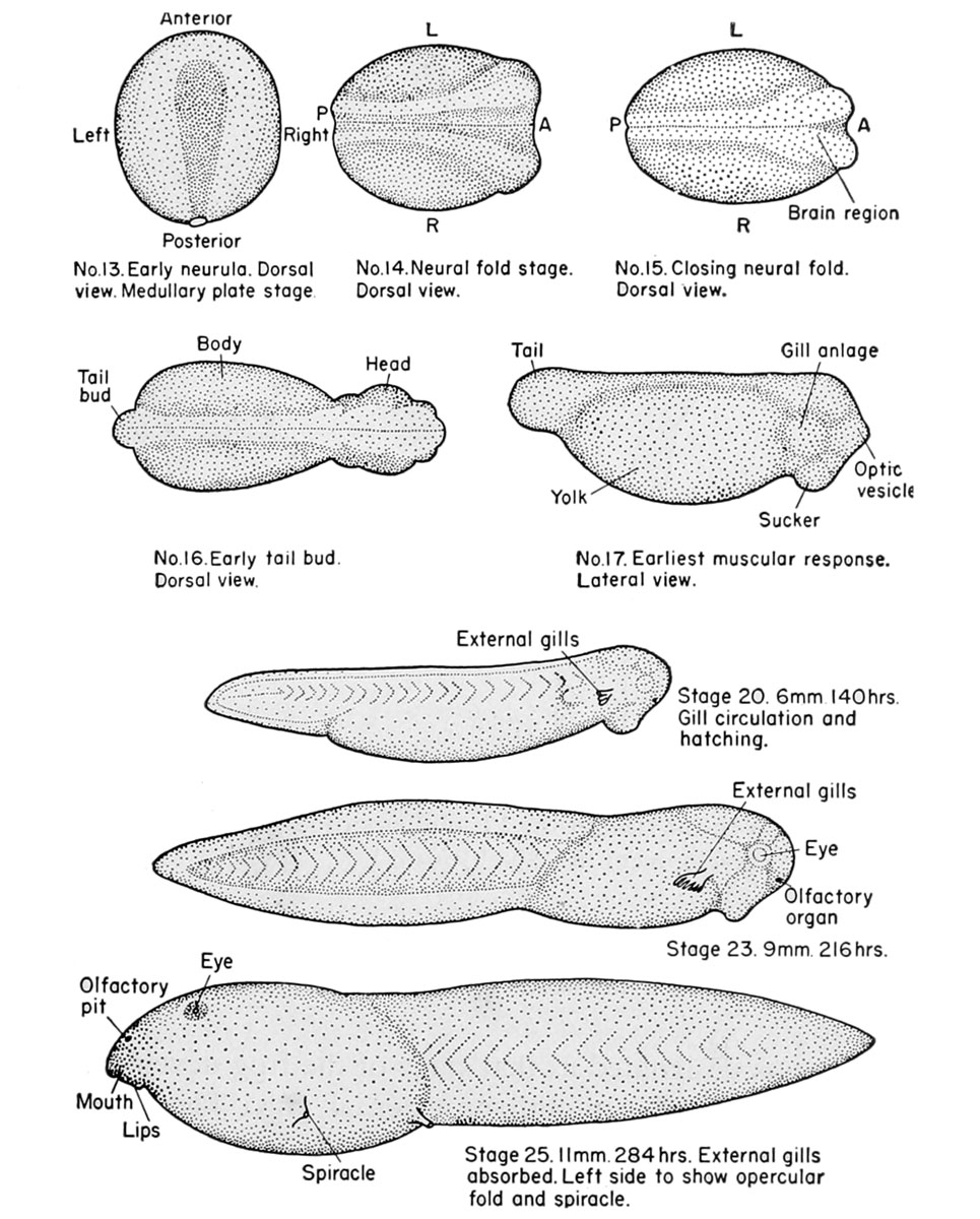
Early development of the frog embryo
(Top) Development of the axial central nervous system.
(Bottom) Development of the external gills and operculum.
No. 13. Early neurula. Dorsal
No. 14. Neural fold stage.
No. 15. Closing neural fold view Medullary plate stage Dorsal view. Dorsal view.
No. 16. Early tail bud. Dorsal view.
No, 17. Earliest musculor response. Lateral view.
Stage 20, 6mm I40hrs Gill circulation and hatching. External gills
Stage 25 11 mm 284hrs External gills Spiracie absorbed. Left side to show opercular fold and spiracle.
| Historic Disclaimer - information about historic embryology pages |
|---|
 |
Reference
Rugh R. Book - The Frog Its Reproduction and Development. (1951) The Blakiston Company.
Cite this page: Hill, M.A. (2024, May 11) Embryology Rugh 007.jpg. Retrieved from https://embryology.med.unsw.edu.au/embryology/index.php/File:Rugh_007.jpg
- © Dr Mark Hill 2024, UNSW Embryology ISBN: 978 0 7334 2609 4 - UNSW CRICOS Provider Code No. 00098G
File history
Click on a date/time to view the file as it appeared at that time.
| Date/Time | Thumbnail | Dimensions | User | Comment | |
|---|---|---|---|---|---|
| current | 14:48, 6 April 2013 | | 965 × 1,200 (220 KB) | Z8600021 (talk | contribs) | ==Early development of the frog embryo== (Top) Development of the axial central nervous system. (Bottom) Development of the external gills and operculum. No. 13. Early neurula. Dorsal No. 14. Neural fold stage. No. 15. Closing neural fold view ... |
You cannot overwrite this file.
File usage
The following 2 pages use this file: