File:Respiratory histology 06.jpg
Respiratory_histology_06.jpg (450 × 600 pixels, file size: 95 KB, MIME type: image/jpeg)
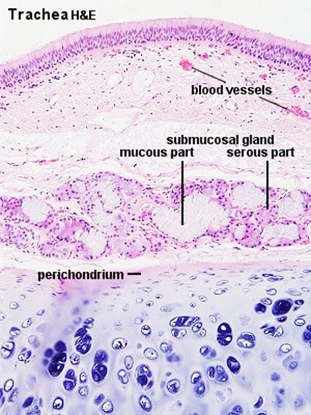
Respiratory Trachea - Glands
Mucosa - formed by epithelium and underlying lamina propria.
- respiratory epithelium - (pseudostratified columnar and ciliated) ciliated cells, goblet cells, brush cells, endocrine cells, surfactant-producing cells (Clara cells), serous cells, basal cells, basement membrane.
- lamina propria - loose connective tissue, many elastic fibres
Submucosa - connective tissue and submucosal glands
Submucosal Glands
(muco-serous) serous (dark) and mucous (light) parts have different staining appearance.
- Mucous secretions - "slimy" (high viscosity) mucous acini cells appear "foamy" or "frothy" and poorly stained (light). nuclei dark and smaller than serous.
- Serous secretions - "watery" (low viscosity) serous acini cell apical cytoplasm is usually well-stained (dark). nuclei round to ovoid located in cell basal cytoplasm.
Cartilage
- perichondrium - surface of cartilage.
- tracheal cartilage - hyaline cartilage, 16 to 20 C-shaped cartilages.
- trachealis muscle - (smooth muscle) Not visible in this section, together with connective tissue fibres, join ends of the cartilages together.
- Trachea Histology Links: Overview HE | Overview VG | Detail 1 HE Detail 2 HE | Respiratory Histology | Histology Stains | Histology
- Respiratory Histology: Bronchiole | Alveolar Duct | Alveoli | EM Alveoli septum | Alveoli Elastin | Trachea 1 | Trachea 2 | labeled lung | unlabeled lung | Respiratory Bronchiole | Lung Reticular Fibres | Nasal Inferior Concha | Nasal Respiratory Epithelium | Olfactory Region overview | Olfactory Region Epithelium | Histology Stains
Links: Histology | Histology Stains | Blue Histology images copyright Lutz Slomianka 1998-2009. The literary and artistic works on the original Blue Histology website may be reproduced, adapted, published and distributed for non-commercial purposes. See also the page Histology Stains.
Cite this page: Hill, M.A. (2024, May 4) Embryology Respiratory histology 06.jpg. Retrieved from https://embryology.med.unsw.edu.au/embryology/index.php/File:Respiratory_histology_06.jpg
- © Dr Mark Hill 2024, UNSW Embryology ISBN: 978 0 7334 2609 4 - UNSW CRICOS Provider Code No. 00098G
File history
Click on a date/time to view the file as it appeared at that time.
| Date/Time | Thumbnail | Dimensions | User | Comment | |
|---|---|---|---|---|---|
| current | 19:54, 27 February 2012 | | 450 × 600 (95 KB) | Z8600021 (talk | contribs) | |
| 15:59, 2 March 2011 |  | 300 × 400 (56 KB) | S8600021 (talk | contribs) | ==Respiratory Trachea== Respiratory histology 06.jpg Original file name: Tra12he.jpg {{Template:Blue Histology}} Category:Respiratory |
You cannot overwrite this file.