File:Radford1908 fig01.jpg
From Embryology
Size of this preview: 800 × 556 pixels. Other resolution: 1,280 × 889 pixels.
Original file (1,280 × 889 pixels, file size: 186 KB, MIME type: image/jpeg)
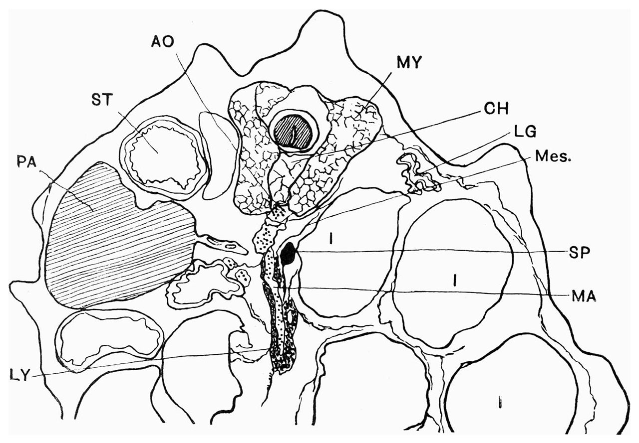
Fig. 1. Transverse section of embryo of 18 mm total length
Showing position of spleen in relation to other organs. Ventral part of section omitted.
AO, aorta; CH, notochord; I, intestine; LG, lung; LY, lymphoid cells; MA,mesenteric artery; Mes., mesentery; MY, myotome ; PA, pancreas; SP, spleen; ST, stomach.
Reference
Radford M. Development of the spleen. (1908) J Anat Physiol. 42: 288-301.
Cite this page: Hill, M.A. (2024, May 6) Embryology Radford1908 fig01.jpg. Retrieved from https://embryology.med.unsw.edu.au/embryology/index.php/File:Radford1908_fig01.jpg
- © Dr Mark Hill 2024, UNSW Embryology ISBN: 978 0 7334 2609 4 - UNSW CRICOS Provider Code No. 00098G
File history
Click on a date/time to view the file as it appeared at that time.
| Date/Time | Thumbnail | Dimensions | User | Comment | |
|---|---|---|---|---|---|
| current | 12:46, 19 July 2019 | | 1,280 × 889 (186 KB) | Z8600021 (talk | contribs) | resize and crop legend |
| 12:42, 19 July 2019 | 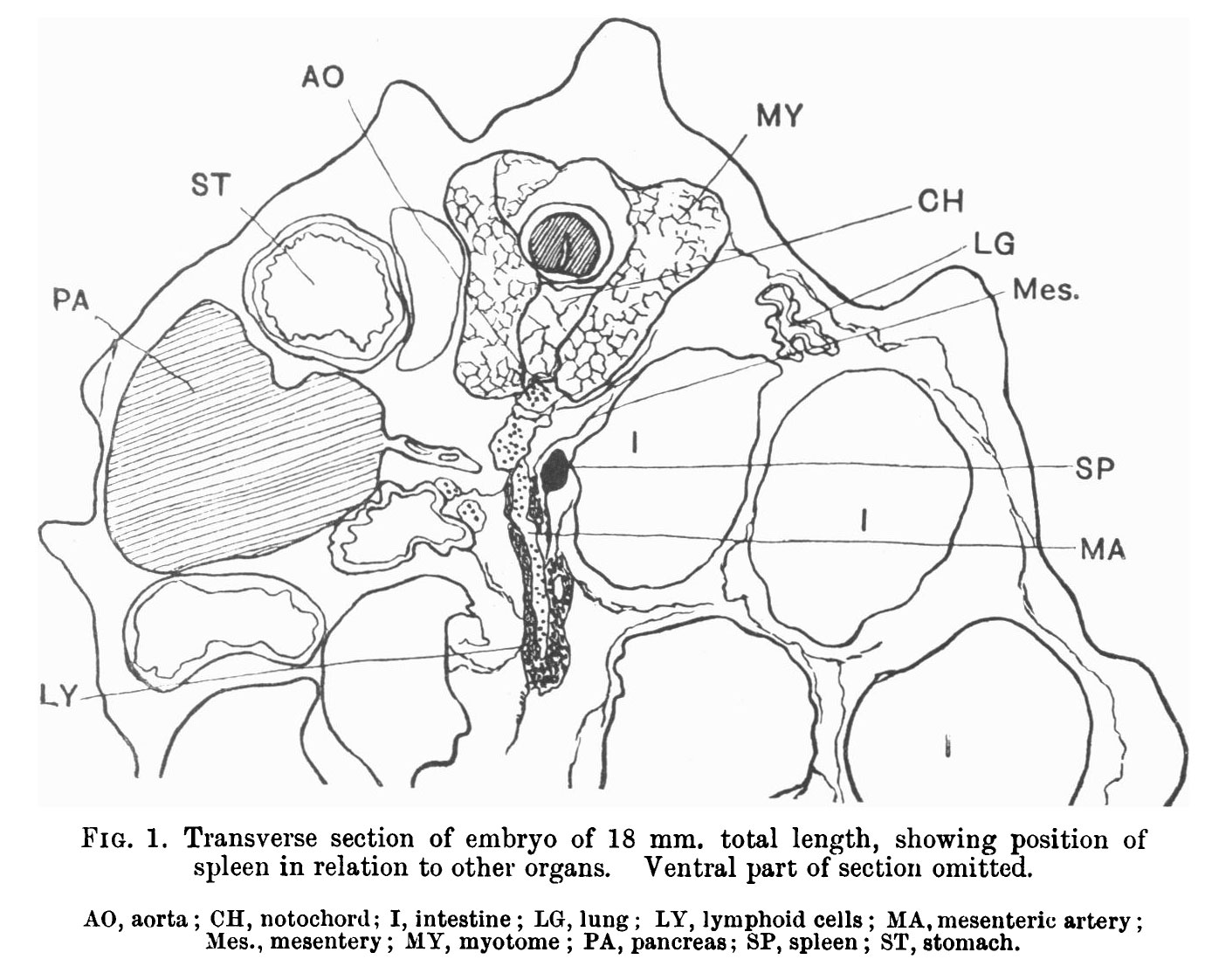 | 1,380 × 1,122 (225 KB) | Z8600021 (talk | contribs) |
You cannot overwrite this file.
File usage
The following page uses this file: