File:Pulmonary Pleura - pseudoglandular and canalicular stages 02.jpg
Original file (671 × 853 pixels, file size: 233 KB, MIME type: image/jpeg)
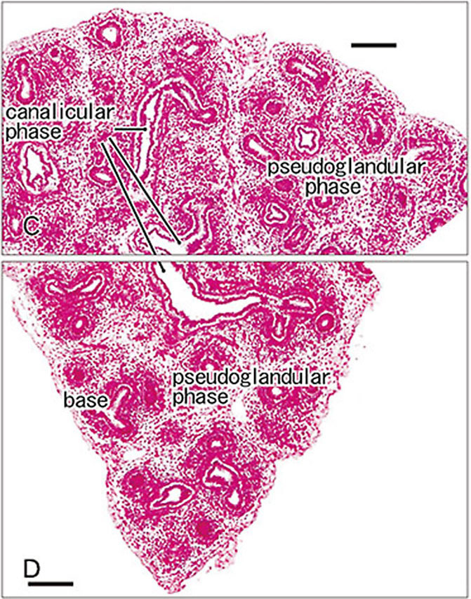
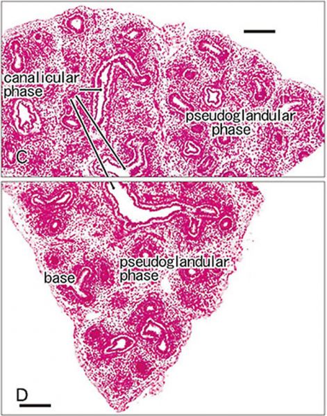
Pulmonary Pleura at the Pseudoglandular and Canalicular Stages
Rough lung surface and pulmonary pleura at the pseudoglandular and canalicular phases. (Stain - Haematoxylin Eosin) Sagittal sections.
Right lung from a specimen at 10 weeks (56 mm CRL) showing canalicular phase of the bronchus was mixed with the pseudoglandular phase. Scale bars = 0.1 mm
Reference
Yamamoto M, Wilting J, Abe H, Murakami G, Rodríguez-Vázquez JF & Abe SI. (2018). Development of the pulmonary pleura with special reference to the lung surface morphology: a study using human fetuses. Anat Cell Biol , 51, 150-157. PMID: 30310706 DOI.
Copyright
Copyright © 2018. Anatomy & Cell Biology
This is an Open Access article distributed under the terms of the Creative Commons Attribution Non-Commercial License (http://creativecommons.org/licenses/by-nc/4.0/) which permits unrestricted non-commercial use, distribution, and reproduction in any medium, provided the original work is properly cited.
Original file name - Fig. 1. Acb-51-150-g001-l.jpg Image adjusted in size and PMID added. Panel D cropped from full figure.
Cite this page: Hill, M.A. (2024, May 16) Embryology Pulmonary Pleura - pseudoglandular and canalicular stages 02.jpg. Retrieved from https://embryology.med.unsw.edu.au/embryology/index.php/File:Pulmonary_Pleura_-_pseudoglandular_and_canalicular_stages_02.jpg
- © Dr Mark Hill 2024, UNSW Embryology ISBN: 978 0 7334 2609 4 - UNSW CRICOS Provider Code No. 00098G
File history
Click on a date/time to view the file as it appeared at that time.
| Date/Time | Thumbnail | Dimensions | User | Comment | |
|---|---|---|---|---|---|
| current | 11:47, 23 February 2019 | | 671 × 853 (233 KB) | Z8600021 (talk | contribs) |
You cannot overwrite this file.
File usage
The following page uses this file: