File:Placental imaging 03.jpg
Original file (869 × 830 pixels, file size: 227 KB, MIME type: image/jpeg)
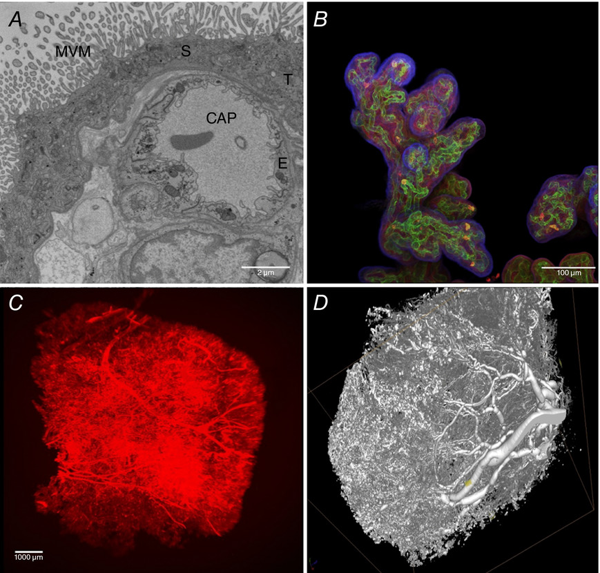
Placental Imaging
A - a transmission electron micrograph of terminal villi showing microvillous membrane (MVM), an underlying capillary (CAP), a syncytiotrophoblast (S), a trophoblast (T) and endothelium (E).
B - projection of an imaged stack (wholemount confocal microscopy), stained with lectin FITC‐AAL for the endothelium (green), rhodamine‐PSA for the stroma (red) and biotin‐DSL for the trophoblast (violet); the DSL was detected with streptavidin 680 and imaging was on a Leica Sp5 confocal microscope, presented as an image stack.
C - villous microcirculation of a term normal placenta perfused with a Ulex Europaeus Agglutinin (UEA) lectin linked to biotin and detected with streptavidin 800.
D - microCT image of a vascular corrosion cast of a term placenta, infused through the umbilical artery with Batson's resin, which was then set, followed by tissue corrosion steps for several days in 20% (w/v) potassium hydroxide.
- Links: placenta | trophoblast | Fig 3A
Reference
Nye GA, Ingram E, Johnstone ED, Jensen OE, Schneider H, Lewis RM, Chernyavsky IL & Brownbill P. (2018). Human placental oxygenation in late gestation: experimental and theoretical approaches. J. Physiol. (Lond.) , 596, 5523-5534. PMID: 29377190 DOI.
Copyright
© 2018 University of Oxford. The Journal of Physiology published by John Wiley & Sons Ltd on behalf of The Physiological Society
This is an open access article under the terms of the Creative Commons Attribution License, which permits use, distribution and reproduction in any medium, provided the original work is properly cited.
Figure 3. Evaluating imaging techniques for use in assessing placental structure Tjp12830-fig-0003-m.jpg
Cite this page: Hill, M.A. (2024, May 21) Embryology Placental imaging 03.jpg. Retrieved from https://embryology.med.unsw.edu.au/embryology/index.php/File:Placental_imaging_03.jpg
- © Dr Mark Hill 2024, UNSW Embryology ISBN: 978 0 7334 2609 4 - UNSW CRICOS Provider Code No. 00098G
File history
Click on a date/time to view the file as it appeared at that time.
| Date/Time | Thumbnail | Dimensions | User | Comment | |
|---|---|---|---|---|---|
| current | 11:11, 10 April 2019 | | 869 × 830 (227 KB) | Z8600021 (talk | contribs) | ==Placental Imaging== A, a transmission electron micrograph of terminal villi showing microvillous membrane (MVM), an underlying capillary (CAP), a syncytiotrophoblast (S), a trophoblast (T) and endothelium (E). B, projection of an imaged stack (whole... |
You cannot overwrite this file.
File usage
The following page uses this file: