File:Placenta histology 001.jpg
From Embryology
Size of this preview: 750 × 600 pixels. Other resolution: 1,280 × 1,024 pixels.
Original file (1,280 × 1,024 pixels, file size: 155 KB, MIME type: image/jpeg)
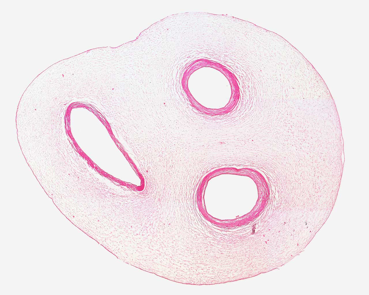
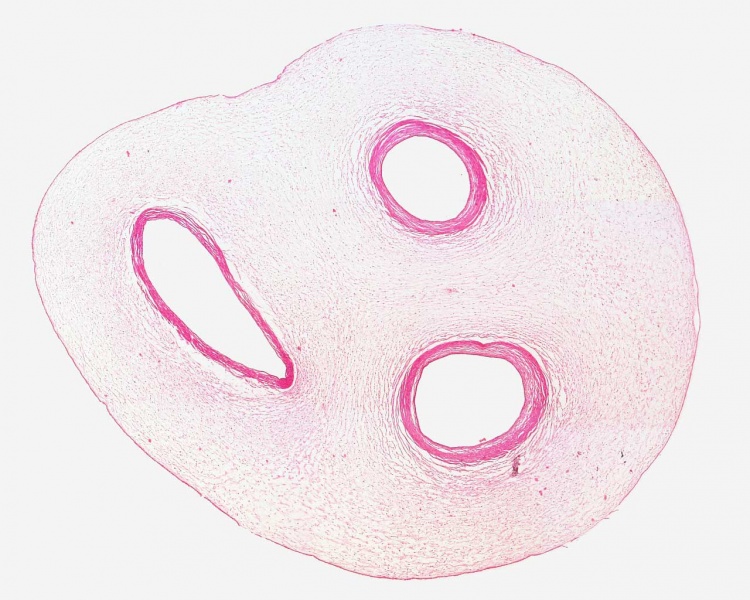
Placenta Cord Histology
- Left - Placental vein
- Right - Paired placental arteries
- Placental Cord Histology: Cord overview | Vein | Artery | Artery | Allantois | Epithelium | Cord overview 1 unlabeled | overview 2 unlabeled | unlabeled vein and connective tissue | unlabeled connective tissue | Villi histology | Placenta Histology
Links: Histology | Histology Stains | Blue Histology images copyright Lutz Slomianka 1998-2009. The literary and artistic works on the original Blue Histology website may be reproduced, adapted, published and distributed for non-commercial purposes. See also the page Histology Stains.
Cite this page: Hill, M.A. (2024, May 4) Embryology Placenta histology 001.jpg. Retrieved from https://embryology.med.unsw.edu.au/embryology/index.php/File:Placenta_histology_001.jpg
- © Dr Mark Hill 2024, UNSW Embryology ISBN: 978 0 7334 2609 4 - UNSW CRICOS Provider Code No. 00098G
File history
Click on a date/time to view the file as it appeared at that time.
| Date/Time | Thumbnail | Dimensions | User | Comment | |
|---|---|---|---|---|---|
| current | 08:01, 31 March 2012 | | 1,280 × 1,024 (155 KB) | Z8600021 (talk | contribs) | |
| 21:21, 23 February 2011 | 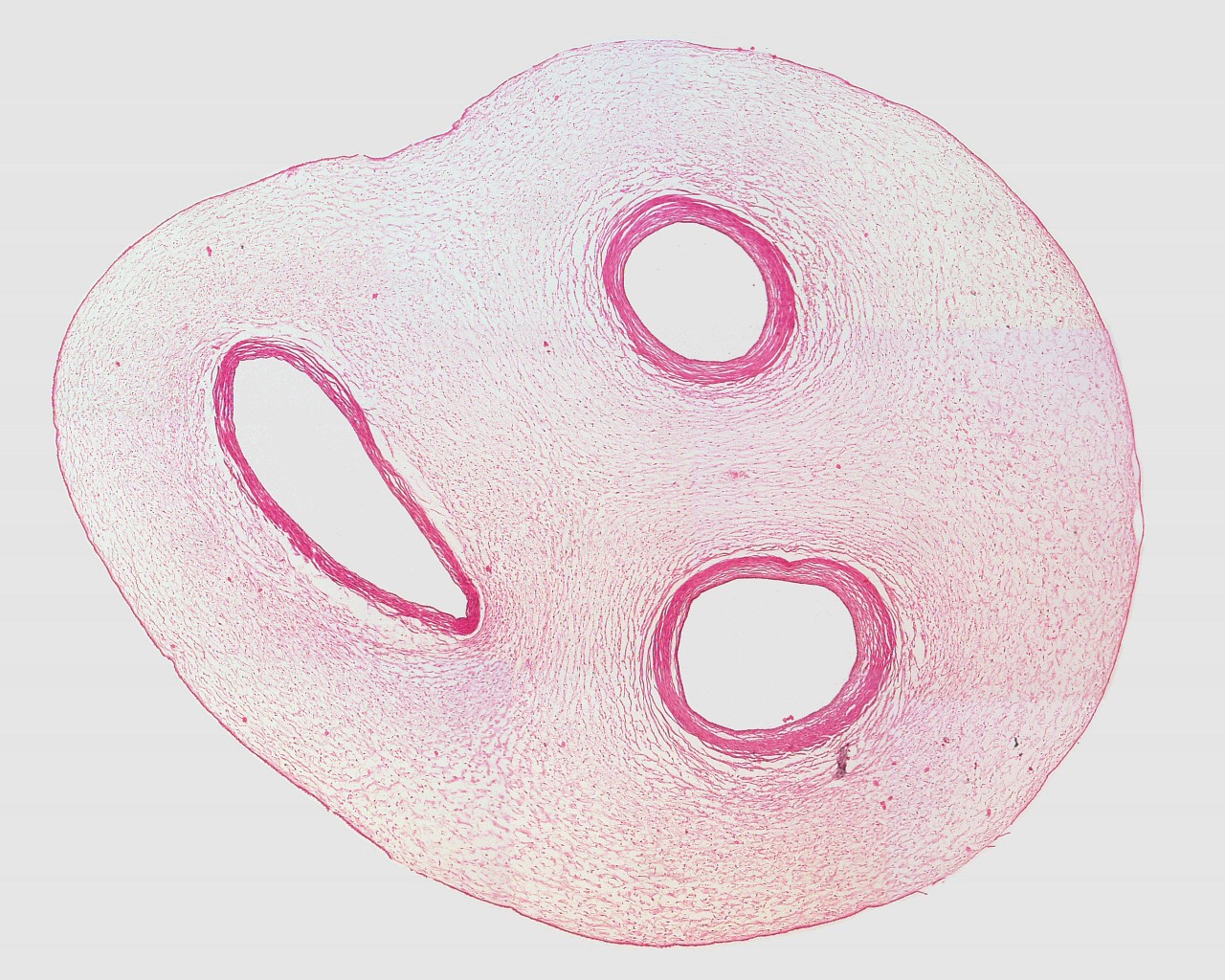 | 1,280 × 1,024 (381 KB) | S8600021 (talk | contribs) | File:Placenta_histology_001.jpg Umbilical_cord,_human_H&E_reproductive_system,_female,_loupe.jpg {{Placenta Histology}} {{Template:Blue Histology}} Category:Placenta Category:Histology |
You cannot overwrite this file.
File usage
The following page uses this file: