File:Patten055.jpg
Patten055.jpg (790 × 567 pixels, file size: 124 KB, MIME type: image/jpeg)
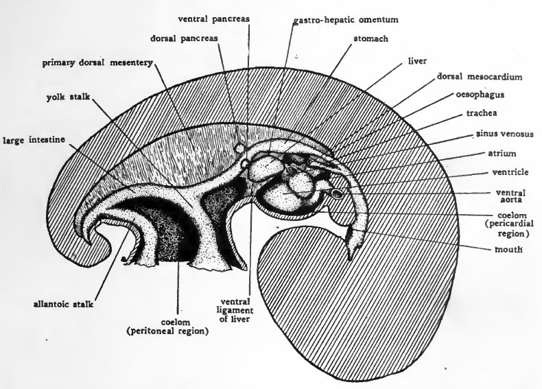
Fig. 55. Schematic lateral view of dissection of four-day chick to show the body cavity and the more important mesenteries
When the ventral mesocardium, and a little later the dorsal mesocardium, breaks through, the primary right and left coelomic chambers become confluent to form the pericardial region of the body cavity (Figs. 24 and 55). Later in development the ventral mesentery farther caudally disappears so that caudally as well as cephahcally an unpaired condition of the coelom is brought about (Fig. 54, H). In the liver region the ventral mesentery does not disappear. The liver arises as an outgrowth from the gut and in its development extends into the ventral mesentery (Fig. 54, G). The portion of the ventral mesentery dorsal to the liver persists as the gastro-hepatic omentum, and the portion ventral to the liver persists as its ventral ligament (falciform ligament) (Fig. 55)
The primary dorsal mesentery persists and forms the supporting membranes of the digestive tube. In the adult its different regions are named according to the parts of the digestive tube with which they are associated, as for example, mesogaster that part of the primary dorsal mesentery which suspends the stomach, mesocolon, that part of the primary dorsal mesentery supporting the colon, etc.
The separation of the body cavity into pericardial, pleural, and peritoneal chambers is accomplished by the formation of septa growing in from the body wall. Consideration of the details of their formation would lead us into stages of development beyond the scope of this book. Those interested in following the later embryology of the chick will find in the appendix references to more exhaustive books, and to a few of the more recent original papers on its development.
- Links: Introduction | Gametes and Fertilization | Segmentation | Entoderm | Primitive Streak and Mesoderm | Primitive Streak to Somites | 24 Hours | 24 to 33 Hours | 33 to 39 Hours | 40 to 50 Hours | Extra-embryonic Membranes | 50 to 55 Hours | Day 3 to 4 | References | All Figures
| Historic Disclaimer - information about historic embryology pages |
|---|
 |
Reference
Patten BM. The Early Embryology of the Chick. (1920) Philadelphia: P. Blakiston's Son and Co.
Cite this page: Hill, M.A. (2024, April 28) Embryology Patten055.jpg. Retrieved from https://embryology.med.unsw.edu.au/embryology/index.php/File:Patten055.jpg
- © Dr Mark Hill 2024, UNSW Embryology ISBN: 978 0 7334 2609 4 - UNSW CRICOS Provider Code No. 00098G
File history
Click on a date/time to view the file as it appeared at that time.
| Date/Time | Thumbnail | Dimensions | User | Comment | |
|---|---|---|---|---|---|
| current | 02:06, 17 January 2011 | | 790 × 567 (124 KB) | S8600021 (talk | contribs) | {{Template:Patten_1920_Figures}} |
You cannot overwrite this file.
File usage
The following 2 pages use this file: