File:Patten001.jpg
From Embryology
Size of this preview: 726 × 600 pixels. Other resolution: 1,002 × 828 pixels.
Original file (1,002 × 828 pixels, file size: 229 KB, MIME type: image/jpeg)
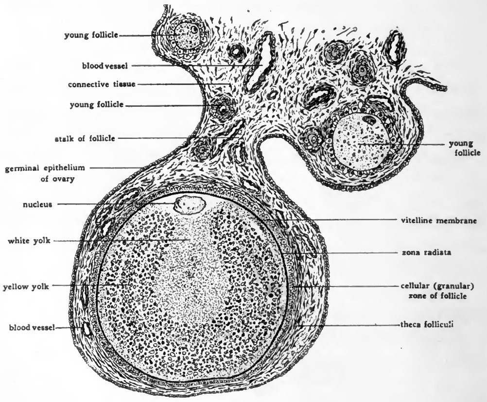
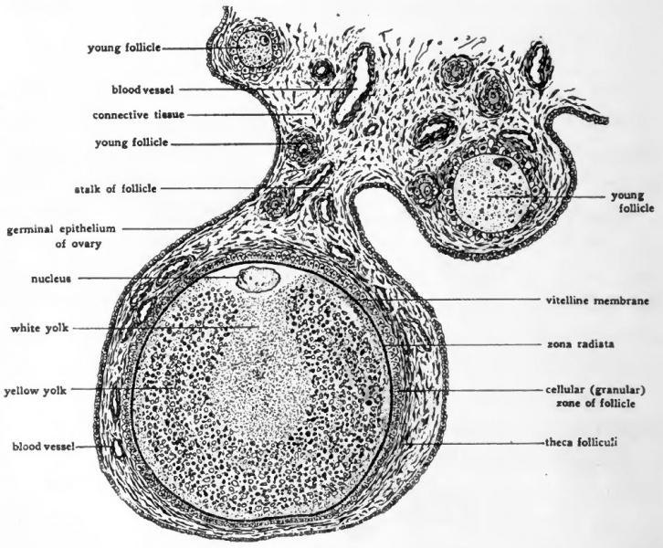
Fig. 1. Diagram showing the structure of a bird ovum still in the ovary
(Modified from Lillie, after Patterson.)
The section shows a follicle containing a nearly mature ovum, together with a small area of the adjacent overian tissue.
- Links: Introduction | Gametes and Fertilization | Segmentation | Entoderm | Primitive Streak and Mesoderm | Primitive Streak to Somites | 24 Hours | 24 to 33 Hours | 33 to 39 Hours | 40 to 50 Hours | Extra-embryonic Membranes | 50 to 55 Hours | Day 3 to 4 | References | All Figures
| Historic Disclaimer - information about historic embryology pages |
|---|
 |
Reference
Patten BM. The Early Embryology of the Chick. (1920) Philadelphia: P. Blakiston's Son and Co.
Cite this page: Hill, M.A. (2024, May 9) Embryology Patten001.jpg. Retrieved from https://embryology.med.unsw.edu.au/embryology/index.php/File:Patten001.jpg
- © Dr Mark Hill 2024, UNSW Embryology ISBN: 978 0 7334 2609 4 - UNSW CRICOS Provider Code No. 00098G
File history
Click on a date/time to view the file as it appeared at that time.
| Date/Time | Thumbnail | Dimensions | User | Comment | |
|---|---|---|---|---|---|
| current | 15:36, 14 January 2011 | | 1,002 × 828 (229 KB) | S8600021 (talk | contribs) | ==Fig. I. Diagram showing the structure of a bird ovum still in the ovary== (Modified from Lillie, after Patterson.) The section shows a follicle containing a nearly mature ovum, together with a small area of the adjacent overian tissue. {{Template:Pa |
You cannot overwrite this file.
File usage
The following 2 pages use this file: