File:Parathyroid position in mouse embryo.jpg
Original file (500 × 641 pixels, file size: 100 KB, MIME type: image/jpeg)
Parathyroid position in mouse embryo
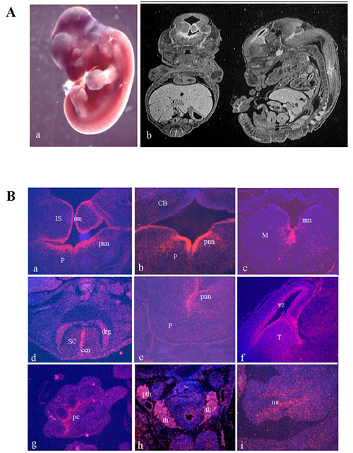
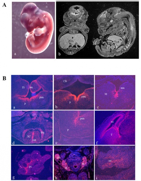
Figure 1. The position of the parathyroid glands are seen in the mouse embryo.
Mig12 expression analysis during embryonic development. (A) Whole mount in situ hybridization on E11.5 mouse embryo showing expression in the central nervous system and in the developing limbs (blue signal, a). Coronal and sagittal sections of E14.5 entire mouse embryos (white signal) (b). (B) Details of coronal (a, b, c, d, h) and sagittal (e, f, g, i) sections of E14.5 mouse embryos. Strong Mig12 expression (red signal) is observed in isthmal (a), pontine (a, b, e) and medulla oblongata (c) neuroepithelia, and it is maintained throughout the entire region of the spinal cord central canal (d). Expression is also observed in dorsal root ganglia (d). Mig12 transcript is detected in the telencephalon at the level of the ventricular zone (f). Signal is also present in other organs: in the perichondrium of the digits (g); in the thyroid (th) and parathyroid (pth) glands (h), and in the phallic part of the urogenital sinus (i). Abbreviations: CB, cerebellum; ccn, central canal neuroepithelium; drg, dorsal root ganglia; IS, isthmus; isn, isthmal neuroepithelium; M, medulla oblongata; mn, medulla oblongata neuroepithelium; P, pons; pc, perichondrium; pnn, pontine neuroepithelium; pth, parathyroid glands; SC, spinal cord; T, telencephalon; th, thyroid gland; us, urogenital sinus; vz, ventricular zone.
Reference
<pubmed>PMC385223</pubmed>
Copyright
© 2004 Berti et al; licensee BioMed Central Ltd. This is an Open Access article: verbatim copying and redistribution of this article are permitted in all media for any purpose, provided this notice is preserved along with the article's original URL.
--Mark Hill (talk) 13:40 7 November 2014 (EST) Assessment - Figure relates to embryonic parathyroid topic and contains reference, copyright and student template. While embryonic development is required to understand what has gone before, you have not included much in fetal development of this gland and its function in the fetus.
- Note - This image was originally uploaded as part of an undergraduate science student project and may contain inaccuracies in either description or acknowledgements. Students have been advised in writing concerning the reuse of content and may accidentally have misunderstood the original terms of use. If image reuse on this non-commercial educational site infringes your existing copyright, please contact the site editor for immediate removal.
File history
Click on a date/time to view the file as it appeared at that time.
| Date/Time | Thumbnail | Dimensions | User | Comment | |
|---|---|---|---|---|---|
| current | 18:04, 8 October 2014 | | 500 × 641 (100 KB) | Z3418837 (talk | contribs) |
You cannot overwrite this file.
File usage
The following 2 pages use this file: