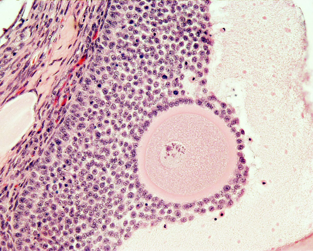
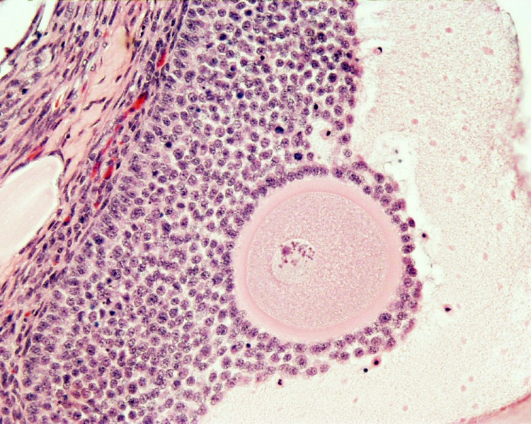
File:Ovary- histology secondary follicle 01.jpg
Original file (1,000 × 800 pixels, file size: 293 KB, MIME type: image/jpeg)
Ovary Antral Follicle with Oocyte
ovary, monkey (Stain - Haematoxylin Eosin)
reproductive system, female, secondary follicle, cumulus oophorus, zona pelucida, granulosa cells, oocyte
- Links: Oocyte Development | Menstrual Cycle | Granulosa cell | Zona pellucida
Links: Histology | Histology Stains | Blue Histology images copyright Lutz Slomianka 1998-2009. The literary and artistic works on the original Blue Histology website may be reproduced, adapted, published and distributed for non-commercial purposes. See also the page Histology Stains.
Cite this page: Hill, M.A. (2024, May 16) Embryology Ovary- histology secondary follicle 01.jpg. Retrieved from https://embryology.med.unsw.edu.au/embryology/index.php/File:Ovary-_histology_secondary_follicle_01.jpg
- © Dr Mark Hill 2024, UNSW Embryology ISBN: 978 0 7334 2609 4 - UNSW CRICOS Provider Code No. 00098G
File history
Click on a date/time to view the file as it appeared at that time.
| Date/Time | Thumbnail | Dimensions | User | Comment | |
|---|---|---|---|---|---|
| current | 23:02, 2 May 2010 | | 1,000 × 800 (293 KB) | S8600021 (talk | contribs) | Ovary Histology (monkey) H&E-reproductive-system,-female,-secondary-follicle,-cumulus-oophorus,-zona-pelucida,-granulosa-cells,-oocyte-x20.jpg Original file name: Image Source: UWA Blue Histology {{Template:Blue Histology}} Category:Ovary [[Cat |
You cannot overwrite this file.
File usage
The following 2 pages use this file: