File:Ovarian tissue extracted after ovarian stimulation.jpeg
From Embryology
Size of this preview: 420 × 599 pixels. Other resolution: 600 × 856 pixels.
Original file (600 × 856 pixels, file size: 345 KB, MIME type: image/jpeg)
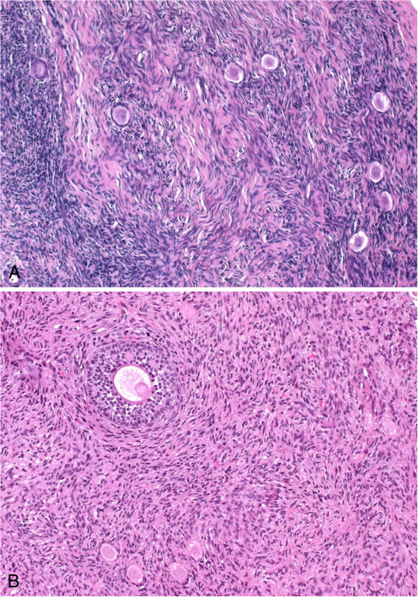
Histological analysis of ovarian tissue extracted immediately after ovarian stimulation.
Ovarian tissue with (A) follicles and (B) one secondary follicle, stained with hematoxylin–eosin (original magnification: × 20).
(Original Figure Legend, PMID 23510640)
Reference
<pubmed>23510640</pubmed>
Copyright
This is an Open Access article distributed under the terms of the Creative Commons Attribution License (http://creativecommons.org/licenses/by/2.0), which permits unrestricted use, distribution, and reproduction in any medium, provided the original work is properly cited.
1477-7827-11-19-1.jpg
- Note - This image was originally uploaded as part of an undergraduate science student project and may contain inaccuracies in either description or acknowledgements. Students have been advised in writing concerning the reuse of content and may accidentally have misunderstood the original terms of use. If image reuse on this non-commercial educational site infringes your existing copyright, please contact the site editor for immediate removal.
File history
Click on a date/time to view the file as it appeared at that time.
| Date/Time | Thumbnail | Dimensions | User | Comment | |
|---|---|---|---|---|---|
| current | 21:25, 9 October 2015 | | 600 × 856 (345 KB) | Z3463667 (talk | contribs) | 1477-7827-11-19-1.jpg PMID 23510640 |
You cannot overwrite this file.
File usage
The following 2 pages use this file: