File:Ovarian follicle growth in vitro.jpg
Original file (1,000 × 769 pixels, file size: 73 KB, MIME type: image/jpeg)
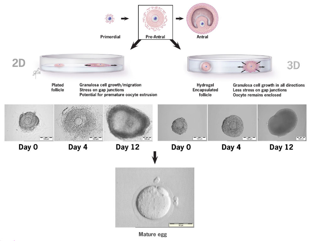
A comparison of follicular growth in a 2-D vs 3-D culture system
2-D culture system
With 2-D growth granulosa cell migration away from the oocyte is evident with time in culture, leaving the oocyte vulnerable for premature extrusion.
3-D culture system
Pre-antral follicles embedded in hydrogel maintain their 3-D architecture. Granulosa cell expansion occurs in all directions resulting in less stress on gap junctions.
Original file name: Figure 1. 1477-7827-8-119-1-l.jpg
Reference
<pubmed>20946661</pubmed>| Reprod Biol Endocrinol.
© 2010 Desai et al; licensee BioMed Central Ltd. This is an Open Access article distributed under the terms of the Creative Commons Attribution License (http://creativecommons.org/licenses/by/2.0), which permits unrestricted use, distribution, and reproduction in any medium, provided the original work is properly cited.
File history
Click on a date/time to view the file as it appeared at that time.
| Date/Time | Thumbnail | Dimensions | User | Comment | |
|---|---|---|---|---|---|
| current | 09:19, 23 February 2011 | | 1,000 × 769 (73 KB) | S8600021 (talk | contribs) | ==A comparison of follicular growth in a 2-D vs 3-D culture system== ===2-D culture system=== With 2-D growth granulosa cell migration away from the oocyte is evident with time in culture, leaving the oocyte vulnerable for premature extrusion. ===3-D |
You cannot overwrite this file.
File usage
The following 2 pages use this file: