File:Oesophagus histology 08.jpg
From Embryology
Size of this preview: 800 × 600 pixels. Other resolution: 1,280 × 960 pixels.
Original file (1,280 × 960 pixels, file size: 353 KB, MIME type: image/jpeg)
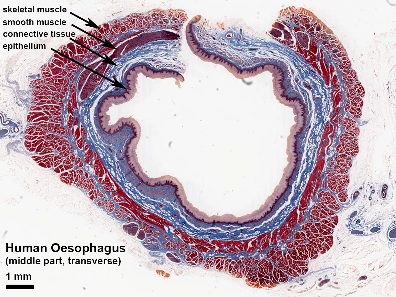
Adult Human Oesophagus
(transverse section)
Middle part showing both skeletal and smooth muscle layers in the wall (Stain - Masson's Trichrome).
- Oesophagus Histology: Skeletal and Smooth Muscle | Submucosa Gland | Muscle | Gland-Muscle Animation | Epithelia and Lamina Propria | Labeled Epithelia | Labeled Connective Tissue | Unlabelled | Unlabelled | Oesophagus Development
Cite this page: Hill, M.A. (2024, May 2) Embryology Oesophagus histology 08.jpg. Retrieved from https://embryology.med.unsw.edu.au/embryology/index.php/File:Oesophagus_histology_08.jpg
- © Dr Mark Hill 2024, UNSW Embryology ISBN: 978 0 7334 2609 4 - UNSW CRICOS Provider Code No. 00098G
File history
Click on a date/time to view the file as it appeared at that time.
| Date/Time | Thumbnail | Dimensions | User | Comment | |
|---|---|---|---|---|---|
| current | 07:02, 3 May 2017 | | 1,280 × 960 (353 KB) | Z8600021 (talk | contribs) | |
| 07:01, 3 May 2017 | 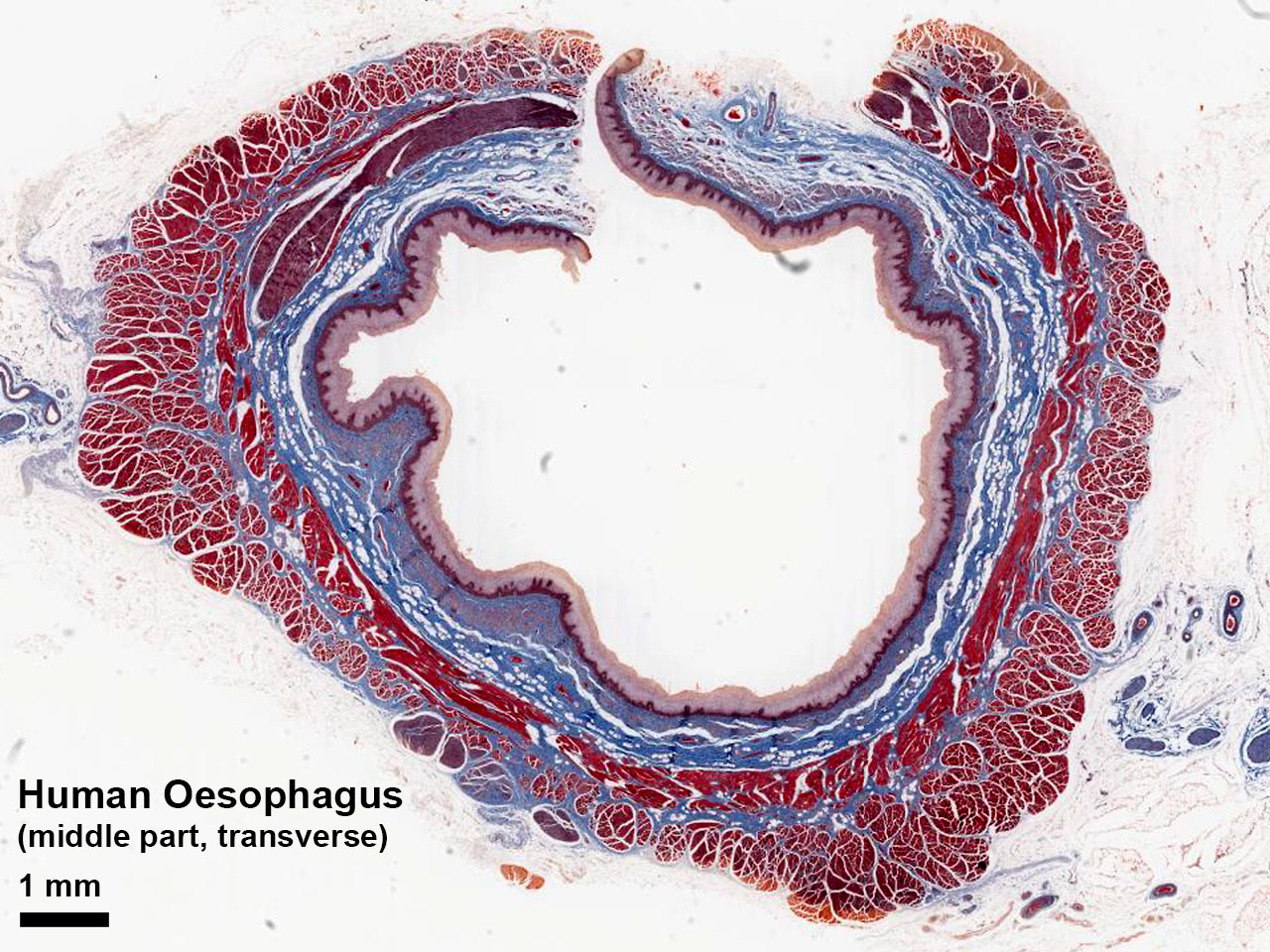 | 1,280 × 960 (343 KB) | Z8600021 (talk | contribs) |
You cannot overwrite this file.
File usage
The following page uses this file: