File:Oesophagus histology 01.jpg
Original file (1,280 × 1,024 pixels, file size: 290 KB, MIME type: image/jpeg)
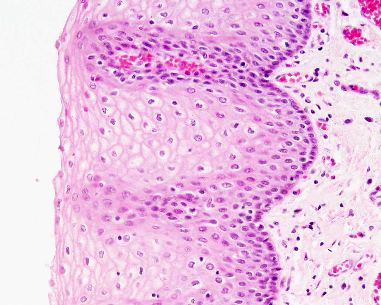
Oesophagus Histology
Unlabelled overview, lumen to the left. See also rotated image lumen to top of image.
- Oesophagus Histology: Skeletal and Smooth Muscle | Submucosa Gland | Muscle | Gland-Muscle Animation | Epithelia and Lamina Propria | Labeled Epithelia | Labeled Connective Tissue | Unlabelled | Unlabelled | Oesophagus Development
Links: Histology | Histology Stains | Blue Histology images copyright Lutz Slomianka 1998-2009. The literary and artistic works on the original Blue Histology website may be reproduced, adapted, published and distributed for non-commercial purposes. See also the page Histology Stains.
Cite this page: Hill, M.A. (2024, May 16) Embryology Oesophagus histology 01.jpg. Retrieved from https://embryology.med.unsw.edu.au/embryology/index.php/File:Oesophagus_histology_01.jpg
- © Dr Mark Hill 2024, UNSW Embryology ISBN: 978 0 7334 2609 4 - UNSW CRICOS Provider Code No. 00098G
File history
Click on a date/time to view the file as it appeared at that time.
| Date/Time | Thumbnail | Dimensions | User | Comment | |
|---|---|---|---|---|---|
| current | 13:16, 5 March 2012 | | 1,280 × 1,024 (290 KB) | Z8600021 (talk | contribs) |
You cannot overwrite this file.
File usage
The following 3 pages use this file: