File:Odgers1939-fig10.jpg
From Embryology

Size of this preview: 290 × 600 pixels. Other resolution: 566 × 1,171 pixels.
Original file (566 × 1,171 pixels, file size: 180 KB, MIME type: image/jpeg)
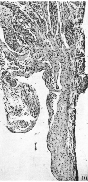
Fig. 10. A section through the right lateral cusp of a full time foetus
( x 60). Note the well-marked annulus fibrosus AR, and that both ventricular muscle, V., and auricular muscle, A., stray on to the base of the cusp to a very small extent.
Reference
Odgers PNB. The development of the atrio-ventricular valves in man. (1939) J Anat. 73: 643-57. PMID 17104787
Cite this page: Hill, M.A. (2024, May 4) Embryology Odgers1939-fig10.jpg. Retrieved from https://embryology.med.unsw.edu.au/embryology/index.php/File:Odgers1939-fig10.jpg
- © Dr Mark Hill 2024, UNSW Embryology ISBN: 978 0 7334 2609 4 - UNSW CRICOS Provider Code No. 00098G
File history
Click on a date/time to view the file as it appeared at that time.
| Date/Time | Thumbnail | Dimensions | User | Comment | |
|---|---|---|---|---|---|
| current | 15:25, 15 November 2015 | | 566 × 1,171 (180 KB) | Z8600021 (talk | contribs) |
You cannot overwrite this file.
File usage
The following 3 pages use this file: