File:Odgers1939-fig09.jpg
From Embryology
Size of this preview: 276 × 600 pixels. Other resolution: 539 × 1,171 pixels.
Original file (539 × 1,171 pixels, file size: 167 KB, MIME type: image/jpeg)
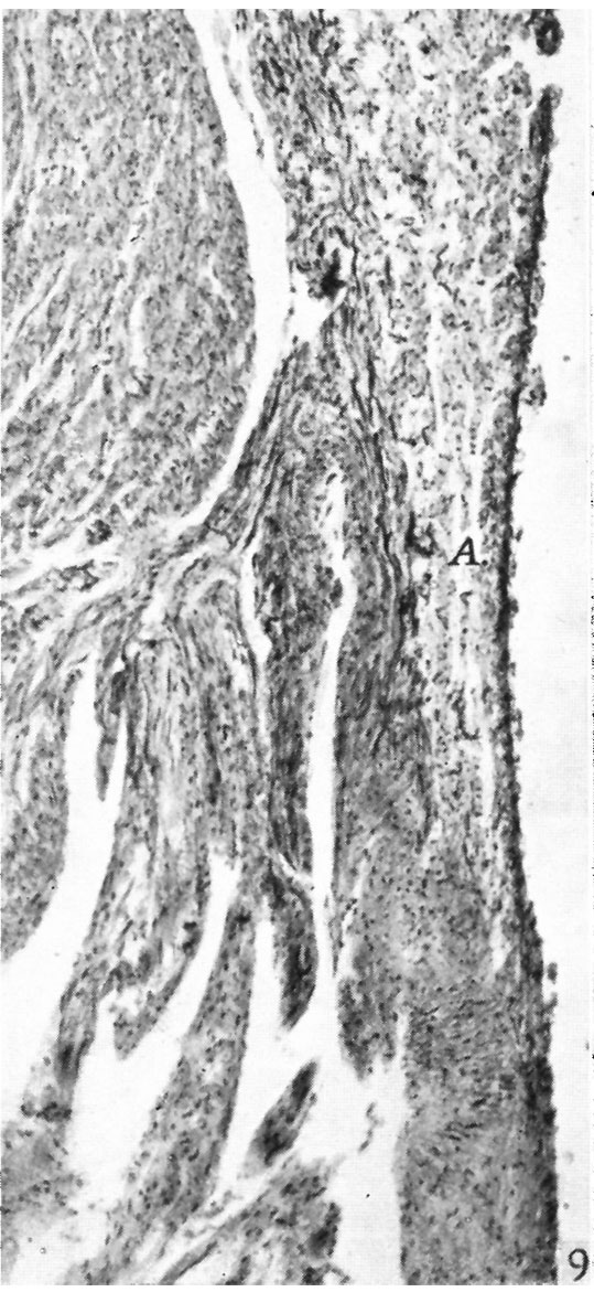
Fig. 9. A section through the right lateral cusp in a 210 mm. foetus
( x 60) to show that auricular muscle, A., does extend on to the base of the cusp in some sections at this stage, the rest of which is entirely collagenous.
Reference
Odgers PNB. The development of the atrio-ventricular valves in man. (1939) J Anat. 73: 643-57. PMID 17104787
Cite this page: Hill, M.A. (2024, May 4) Embryology Odgers1939-fig09.jpg. Retrieved from https://embryology.med.unsw.edu.au/embryology/index.php/File:Odgers1939-fig09.jpg
- © Dr Mark Hill 2024, UNSW Embryology ISBN: 978 0 7334 2609 4 - UNSW CRICOS Provider Code No. 00098G
File history
Click on a date/time to view the file as it appeared at that time.
| Date/Time | Thumbnail | Dimensions | User | Comment | |
|---|---|---|---|---|---|
| current | 15:25, 15 November 2015 | | 539 × 1,171 (167 KB) | Z8600021 (talk | contribs) |
You cannot overwrite this file.
File usage
The following 3 pages use this file: