File:Odgers1939-fig08.jpg

Original file (557 × 1,171 pixels, file size: 176 KB, MIME type: image/jpeg)
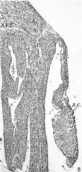
Fig. 8. A section through the right lateral cusp in an 85 mm. foetus
( x 60). The cusp is now collagenous and is continuous with the A.-V. sulcus, A.V.S., this continuity separating auricrular muscle, A., from that of the ventricle, V. Ventricular muscle just comes over on to the base of the cusp. R.C'. marks the apparent remains of cushion tissue.
Reference
Odgers PNB. The development of the atrio-ventricular valves in man. (1939) J Anat. 73: 643-57. PMID 17104787
Cite this page: Hill, M.A. (2024, May 23) Embryology Odgers1939-fig08.jpg. Retrieved from https://embryology.med.unsw.edu.au/embryology/index.php/File:Odgers1939-fig08.jpg
- © Dr Mark Hill 2024, UNSW Embryology ISBN: 978 0 7334 2609 4 - UNSW CRICOS Provider Code No. 00098G
File history
Click on a date/time to view the file as it appeared at that time.
| Date/Time | Thumbnail | Dimensions | User | Comment | |
|---|---|---|---|---|---|
| current | 15:25, 15 November 2015 | | 557 × 1,171 (176 KB) | Z8600021 (talk | contribs) |
You cannot overwrite this file.
File usage
The following 3 pages use this file: