File:Odgers1939-fig02.jpg
From Embryology

Size of this preview: 351 × 600 pixels. Other resolution: 523 × 894 pixels.
Original file (523 × 894 pixels, file size: 157 KB, MIME type: image/jpeg)
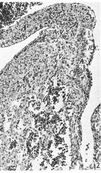
Fig. 2. A section of the right lateral cusp in a 17-5 mm. embryo
( x 109) to show the angulation of the right A.-V. sulcus, A. V.S., which is still separated from the right lateral cushion, R.0., by the junction of auricular, A., with ventricular muscle, V.
File history
Click on a date/time to view the file as it appeared at that time.
| Date/Time | Thumbnail | Dimensions | User | Comment | |
|---|---|---|---|---|---|
| current | 15:00, 15 November 2015 | | 523 × 894 (157 KB) | Z8600021 (talk | contribs) |
You cannot overwrite this file.
File usage
The following 3 pages use this file: