File:Odgers1935 plate01.jpg
Original file (1,280 × 1,459 pixels, file size: 300 KB, MIME type: image/jpeg)
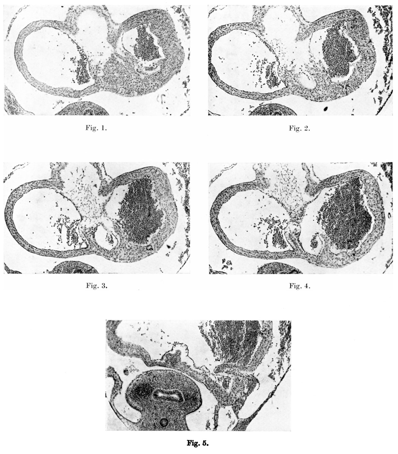
Plate I
Figs. 1-4. These microphotographs of sections of the aurieles in a 5-mm. embryo ( x 60) show in fig. I the myocardium spreading into the subendocardial space from the dorsal wall to form the septum primum. Figs. 2-4 are photographs of alternate sections next below this and in them a lacuna appears in the myocardial proliferation, ultimately splitting this into two. The left limb forms the septum primum, the right limb the left venous valve, the connection between the two persisting as the septum spurium.
Fig. 5 is a microphotograph of the same heart 11 sections below the last. This shows the right valve as being formed by the coaptation of the anterior wall of the right horn of the sinus venosus with the posterior wall of the auricle. The left venous valve is seen as a small projection immediately to the left and slightly anterior to the right valve.
Reference
Odgers PNB. The formation of the venous valves, the foramen secundum and the septum secundum in the human heart. (1935) J. Anat., 69: 412-422. PMID 17104548
Cite this page: Hill, M.A. (2024, May 21) Embryology Odgers1935 plate01.jpg. Retrieved from https://embryology.med.unsw.edu.au/embryology/index.php/File:Odgers1935_plate01.jpg
- © Dr Mark Hill 2024, UNSW Embryology ISBN: 978 0 7334 2609 4 - UNSW CRICOS Provider Code No. 00098G
File history
Click on a date/time to view the file as it appeared at that time.
| Date/Time | Thumbnail | Dimensions | User | Comment | |
|---|---|---|---|---|---|
| current | 14:53, 28 February 2017 | | 1,280 × 1,459 (300 KB) | Z8600021 (talk | contribs) | |
| 14:53, 28 February 2017 | 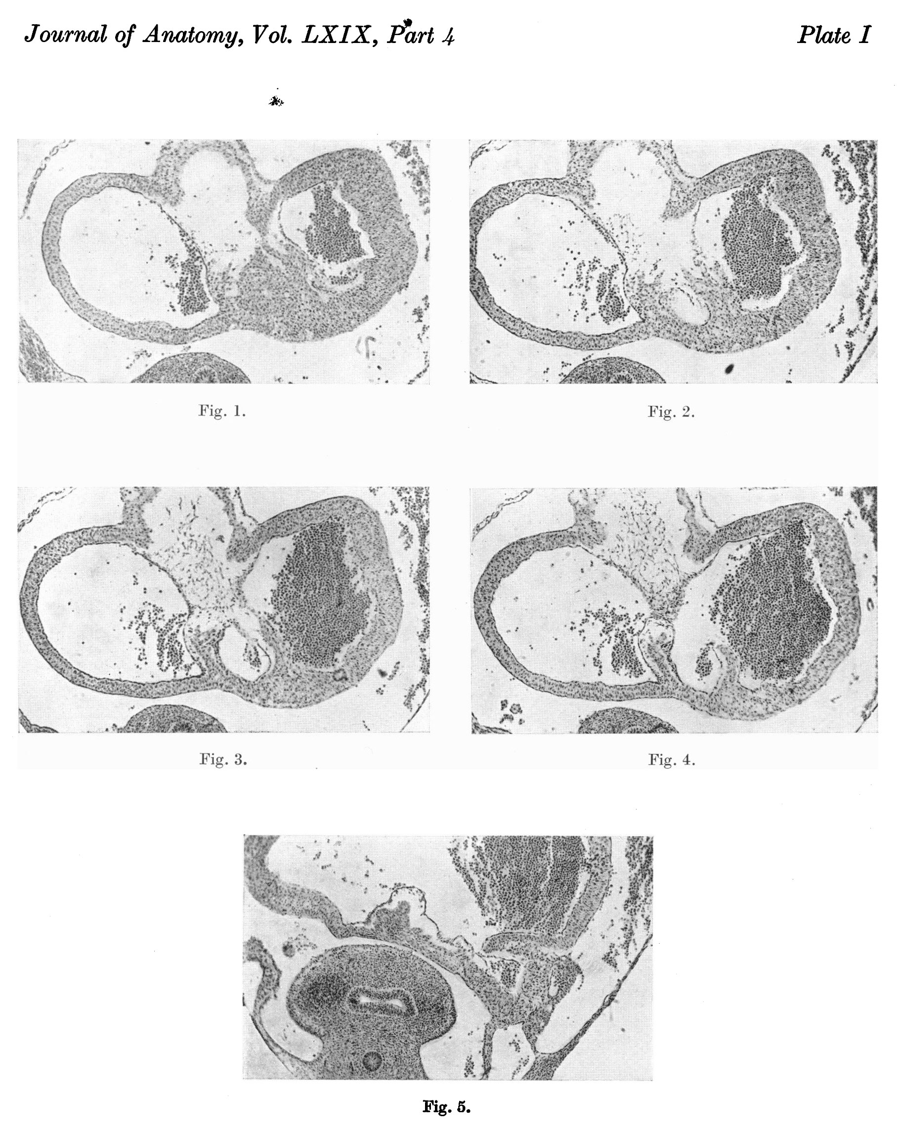 | 1,815 × 2,288 (871 KB) | Z8600021 (talk | contribs) |
You cannot overwrite this file.
File usage
The following page uses this file: