File:Newborn - vernix caseosa.jpg
Newborn_-_vernix_caseosa.jpg (800 × 463 pixels, file size: 73 KB, MIME type: image/jpeg)
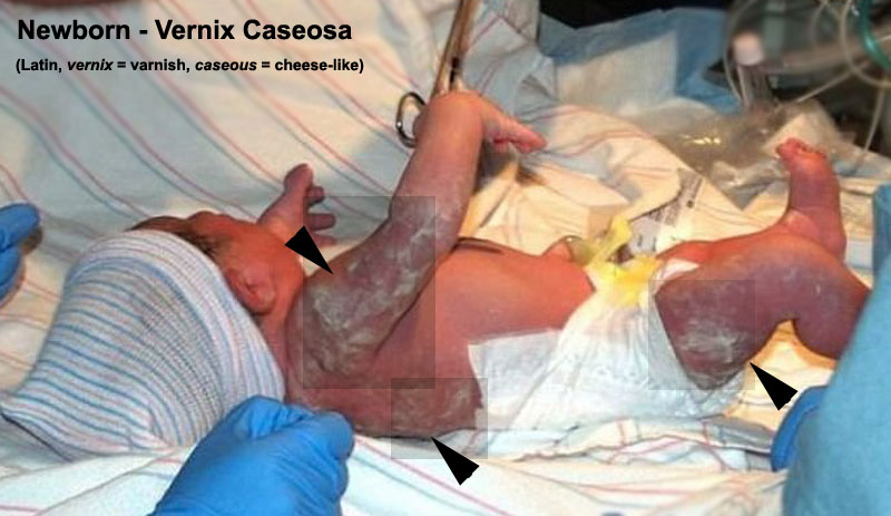
Newborn - Vernix Caseosa
The white material in shaded boxes and arrowed is part of the vernix caseosa that has not yet been removed.
Vernix caseosa forms on the fetus by the developing epidermis shedding the periderm cell layer and then mixed with sebaceous gland sebum secretions within the epithelial walls. Vernix cases begins to cover the fetal skin from about week 17 (GA week 19).
Vernix components - water (81%), lipid (9%), and proteins (10%)
Reference
JazlynRoseVernixByPhilKonstantin.jpg http://en.wikipedia.org/wiki/File:JazlynRoseVernixByPhilKonstantin.jpg (modified in size and labeling)
Cite this page: Hill, M.A. (2024, May 4) Embryology Newborn - vernix caseosa.jpg. Retrieved from https://embryology.med.unsw.edu.au/embryology/index.php/File:Newborn_-_vernix_caseosa.jpg
- © Dr Mark Hill 2024, UNSW Embryology ISBN: 978 0 7334 2609 4 - UNSW CRICOS Provider Code No. 00098G
File history
Click on a date/time to view the file as it appeared at that time.
| Date/Time | Thumbnail | Dimensions | User | Comment | |
|---|---|---|---|---|---|
| current | 14:49, 13 October 2010 | | 800 × 463 (73 KB) | S8600021 (talk | contribs) | ==Newborn - Vernix Caseosa== The white material in shaded boxes and arrowed is part of the vernix that has not yet been removed. Image source: JazlynRoseVernixByPhilKonstantin.jpg http://en.wikipedia.org/wiki/File:JazlynRoseVernixByPhilKonstantin.jp |
You cannot overwrite this file.