File:Mouse ventral body wall development 02.jpg

Original file (1,000 × 1,222 pixels, file size: 214 KB, MIME type: image/jpeg)
Mouse Ventral Body Wall Development
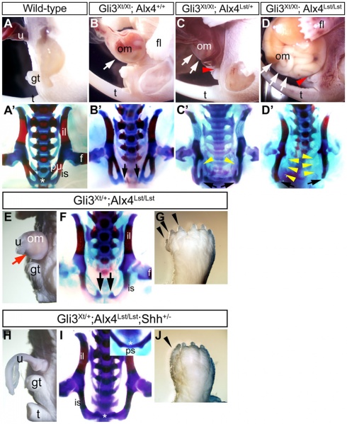
The omphalocele, pubic diastasis, loss of pubic bones and polydactyly in Gli3Xt; Alx4Lst; Shh combinatorial mutants.
The lateral view of the embryonic ventral body wall (A–D, E, H) and frontal view of the pelvic girdle (A'–D', F, I). Gli3Xt/Xt embryos (B), Gli3Xt/Xt; Alx4Lst/+ embryos (C) and Gli3Xt/Xt; Alx4Lst/Lst embryos (D) showed a graded extent of omphaloceles by the introduction of additional Alx4Lst alleles into the Gli3Xt/Xt background (B–D; white arrows). The dorsal parts of the genital tubercle were hypoplastic in Gli3Xt/Xt; Alx4Lst/+ and Gli3Xt/Xt; Alx4Lst/Lst embryos (C, D; red arrowheads).
The pubic symphysis of wild-type embryo was already formed at E18.5 (A'; asterisk). Pubic diastasis also became evident by the introduction of Alx4Lst mutation (B'–D'). Gli3Xt/Xt; Alx4Lst/+ and Gli3Xt/Xt; Alx4Lst/Lst embryos showed partial loss of pubic bone components (C', D'; yellow arrowheads). Black arrows indicate the unclosed pelvis. Gli3Xt/+; Alx4Lst/Lst embryos showed omphalocele (E; red arrow), severe polydactyly (G), pubic diastasis and loss of pubic bones (F). Black arrows show the unclosed pelvis. Gli3Xt/+; Alx4Lst/Lst; Shh+/− embryos did not show omphalocele phenotypes (H). The pubic symphysis was formed but pubic bones were lost (I; asterisk). Polydactyly was still observed in Gli3Xt/+; Alx4Lst/Lst; Shh+/− embryos (J). Black arrowheads indicate extra digits.
- f - femur
- fl - fore limb
- gt - genital tubercle
- il - iliac bone
- is - ischial bone
- om - omphalocele
- ps - pubic symphysis
- pu - pubic bone
- t - tail
- uc - umbilical cord
Original file name: Figure 3. Journal.pone.0016260.g003.jpg doi:10.1371/journal.pone.0016260.g003
Reference
Matsumaru D, Haraguchi R, Miyagawa S, Motoyama J, Nakagata N, Meijlink F & Yamada G. (2011). Genetic analysis of Hedgehog signaling in ventral body wall development and the onset of omphalocele formation. PLoS ONE , 6, e16260. PMID: 21283718 DOI.
Copyright
© 2011 Matsumaru et al. This is an open-access article distributed under the terms of the Creative Commons Attribution License, which permits unrestricted use, distribution, and reproduction in any medium, provided the original author and source are credited.
Cite this page: Hill, M.A. (2024, May 11) Embryology Mouse ventral body wall development 02.jpg. Retrieved from https://embryology.med.unsw.edu.au/embryology/index.php/File:Mouse_ventral_body_wall_development_02.jpg
- © Dr Mark Hill 2024, UNSW Embryology ISBN: 978 0 7334 2609 4 - UNSW CRICOS Provider Code No. 00098G
File history
Click on a date/time to view the file as it appeared at that time.
| Date/Time | Thumbnail | Dimensions | User | Comment | |
|---|---|---|---|---|---|
| current | 16:04, 2 September 2011 | | 1,000 × 1,222 (214 KB) | S8600021 (talk | contribs) | ==Mouse Ventral Body Wall Development== The omphalocele, pubic diastasis, loss of pubic bones and polydactyly in Gli3Xt; Alx4Lst; Shh combinatorial mutants. The lateral view of the embryonic ventral body wall (A–D, E, H) and frontal view of the pelvic |
You cannot overwrite this file.
File usage
The following page uses this file: