File:Mouse-lower incisor teeth.jpg
Original file (1,000 × 497 pixels, file size: 85 KB, MIME type: image/jpeg)
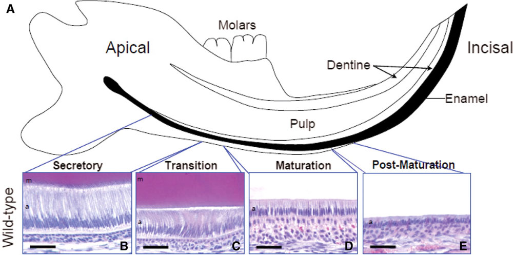
Histological analysis of the lower incisor teeth.
(A) Diagrammatic representation of the mouse lower incisor tooth within the mandible. As indicated in black, enamel production commences at the apical region of the tooth and proceeds towards the incisal edge.
(B–E) The secretory, transition, maturation and post-maturation stages of amelogenesis showed characteristically normal morphology in the WT lower incisor tooth.
Extract from Figure 4. http://hmg.oxfordjournals.org/content/vol19/issue7/images/large/ddq00104.jpeg
A mutation in the mouse Amelx tri-tyrosyl domain results in impaired secretion of amelogenin and phenocopies human X-linked amelogenesis imperfecta. Barron MJ, Brookes SJ, Kirkham J, Shore RC, Hunt C, Mironov A, Kingswell NJ, Maycock J, Shuttleworth CA, Dixon MJ. Hum Mol Genet. 2010 Apr 1;19(7):1230-47. Epub 2010 Jan 12. PMID: 20067920 Hum Mol Genet.
© The Author 2010. Published by Oxford University Press
This is an Open Access article distributed under the terms of the Creative Commons Attribution Non-Commercial License (http://creativecommons.org/licenses/by-nc/2.5/uk) which permits unrestricted non-commercial use, distribution, and reproduction in any medium, provided the original work is properly cited.
File history
Click on a date/time to view the file as it appeared at that time.
| Date/Time | Thumbnail | Dimensions | User | Comment | |
|---|---|---|---|---|---|
| current | 12:31, 22 April 2010 | | 1,000 × 497 (85 KB) | S8600021 (talk | contribs) | Histological analysis of the lower incisor teeth. (A) Diagrammatic representation of the mouse lower incisor tooth within the mandible. As indicated in black, enamel production commences at the apical region of the tooth and proceeds towards the incisal |
You cannot overwrite this file.
File usage
The following page uses this file: