File:Monocyte EM01.jpg
Original file (923 × 1,000 pixels, file size: 221 KB, MIME type: image/jpeg)
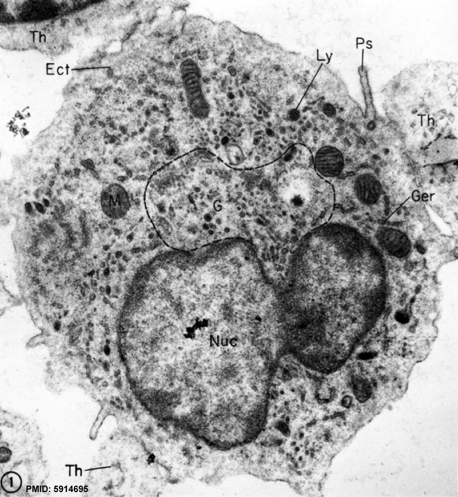
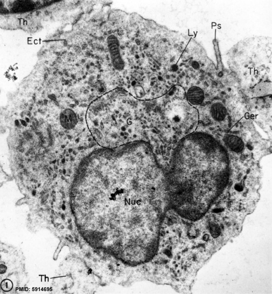
Monocyte Electron Micrograph
This electron micrograph illustrates the salient features of a circulating monocyte.
- Ly - lysosomal granules variably density.
- G - Golgi zone is small, but well developed; its external limits are demarcated by the dotted line.
- Get - Rough-surfaced ER (rough endoplasmic reticulum), many channels of which are dilated by electron-opaque secretory material, is abundant.
- M - Mitochondria are distributed randomly.
- Ps - pseudopodia. A narrow but distinct zone of ectoplasm (Ect) rings the cell and continues into pseudopodia.
- Th - thrombocytes.
X 22,500.
Reference
<pubmed>5914695</pubmed>
Copyright
Rockefeller University Press - Copyright Policy This article is distributed under the terms of an Attribution–Noncommercial–Share Alike–No Mirror Sites license for the first six months after the publication date (see http://www.jcb.org/misc/terms.shtml). After six months it is available under a Creative Commons License (Attribution–Noncommercial–Share Alike 4.0 Unported license, as described at https://creativecommons.org/licenses/by-nc-sa/4.0/ ). (More? Help:Copyright Tutorial)
File history
Click on a date/time to view the file as it appeared at that time.
| Date/Time | Thumbnail | Dimensions | User | Comment | |
|---|---|---|---|---|---|
| current | 13:28, 4 March 2012 | | 923 × 1,000 (221 KB) | Z8600021 (talk | contribs) | ==Monocyte== This electron micrograph illustrates the salient features of a circulating monocyte. * A myriad of variably dense lysosomal granules (Ly) is present. * The Golgi zone (G) is small, but well developed; its external limits are demarcated by |
You cannot overwrite this file.
File usage
There are no pages that use this file.