File:Model embryo to 32 cell stage 001.jpg
From Embryology
No higher resolution available.
Model_embryo_to_32_cell_stage_001.jpg (696 × 256 pixels, file size: 22 KB, MIME type: image/jpeg)
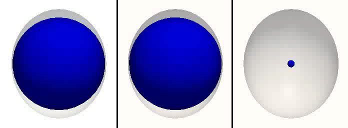
Model Embryo to 32 cell stage
First image in the simulation of embryonic development up to 32 cell stage.
- First pane - CDX2 levels in cells.
- Second pane - inner and outer cell status as well as polarization directions.
- Third pane - nuclei with different colors representing lineage of four cell embryo.
Reference
<pubmed>21573197</pubmed>| PMC3088645 | PLoS Comput Biol.
© 2011 Krupinski et al. This is an open-access article distributed under the terms of the Creative Commons Attribution License, which permits unrestricted use, distribution, and reproduction in any medium, provided the original author and source are credited.
Original file name: Video 1 journal.pcbi.1001128.s012.avi (Converted using Quicktime 7) [[Category:Cartoon]
File history
Click on a date/time to view the file as it appeared at that time.
| Date/Time | Thumbnail | Dimensions | User | Comment | |
|---|---|---|---|---|---|
| current | 10:07, 11 November 2011 | 696 × 256 (22 KB) | S8600021 (talk | contribs) | ==Model Embryo to 32 cell stage== Simulation of embryonic development up to 32 cell stage. * First pane - CDX2 levels in cells. * Second pane - inner and outer cell status as well as polarization directions. * Third pane - nuclei with different color |
You cannot overwrite this file.
File usage
The following 3 pages use this file: