File:Minot1897 003.jpg
From Embryology
Size of this preview: 800 × 482 pixels. Other resolution: 1,176 × 708 pixels.
Original file (1,176 × 708 pixels, file size: 374 KB, MIME type: image/jpeg)
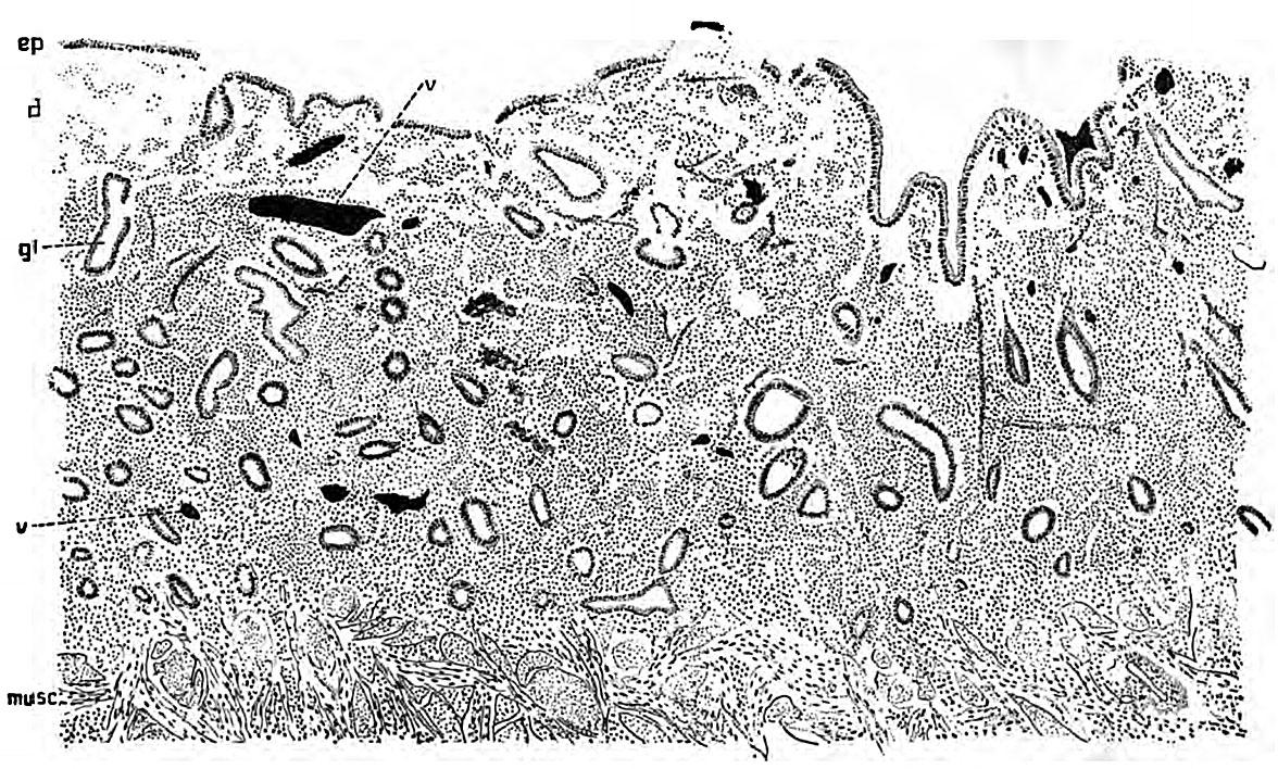
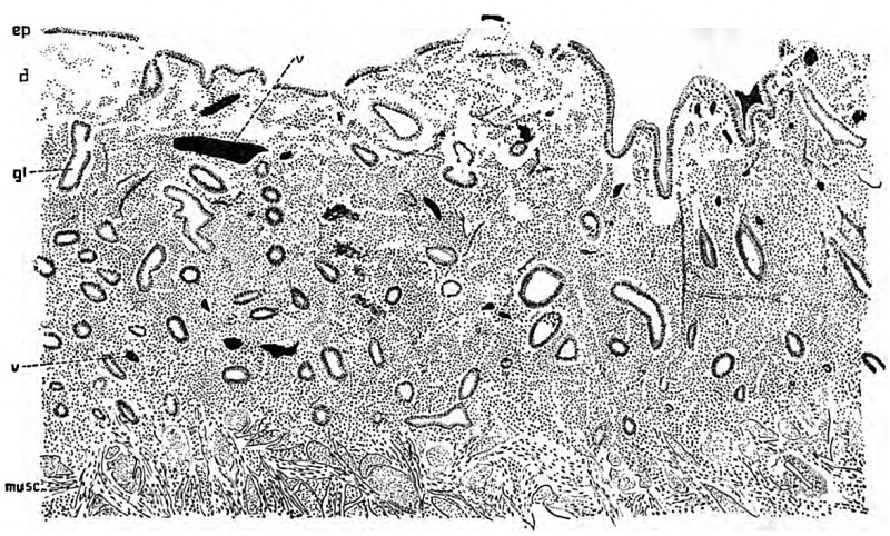
Fig. 3. Mucous membrane of a virgin uterus during the first day of menstruation
| Historic Disclaimer - information about historic embryology pages |
|---|
 |
Reference
Minot CS. Human Embryology. (1897) London: The Macmillan Company.
Cite this page: Hill, M.A. (2024, May 19) Embryology Minot1897 003.jpg. Retrieved from https://embryology.med.unsw.edu.au/embryology/index.php/File:Minot1897_003.jpg
- © Dr Mark Hill 2024, UNSW Embryology ISBN: 978 0 7334 2609 4 - UNSW CRICOS Provider Code No. 00098G
File history
Click on a date/time to view the file as it appeared at that time.
| Date/Time | Thumbnail | Dimensions | User | Comment | |
|---|---|---|---|---|---|
| current | 08:01, 18 June 2015 | | 1,176 × 708 (374 KB) | Z8600021 (talk | contribs) | |
| 08:00, 18 June 2015 | 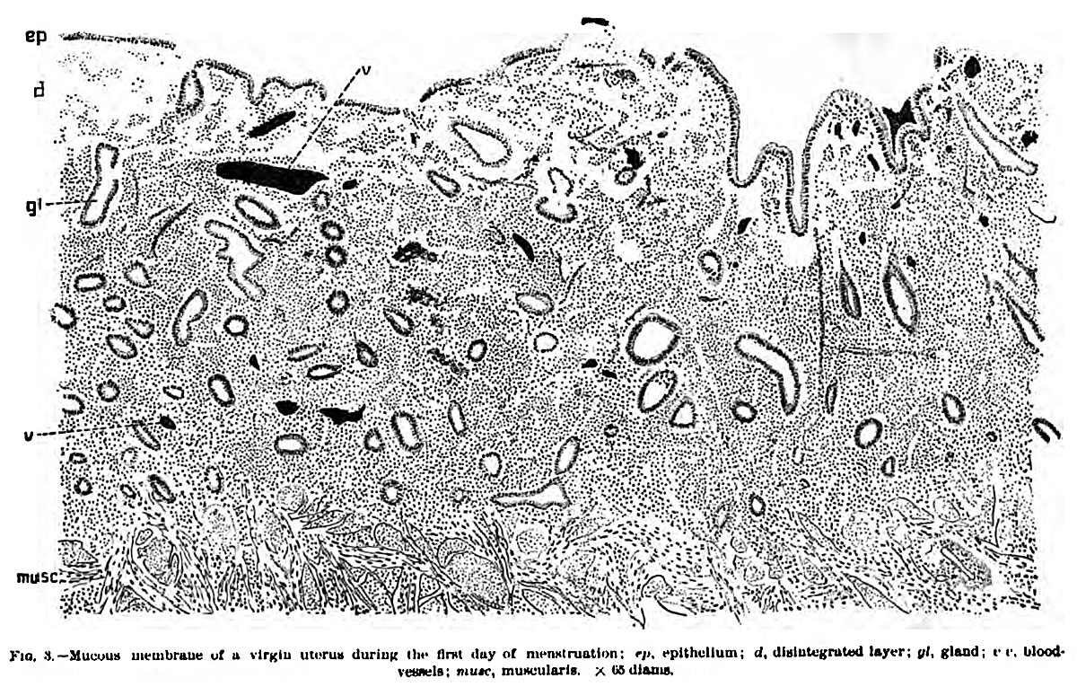 | 1,200 × 765 (390 KB) | Z8600021 (talk | contribs) | ==Fig. 8. Mucous membrane of a virgin uterus during the first day of menstruation== {{Minot1897 figures}} Category:Uterus |
You cannot overwrite this file.
File usage
The following 2 pages use this file: