File:Mammary anatomy.jpg
From Embryology
Size of this preview: 512 × 599 pixels. Other resolution: 600 × 702 pixels.
Original file (600 × 702 pixels, file size: 58 KB, MIME type: image/jpeg)
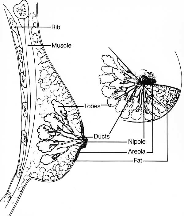
Human Mammary Anatomy
Cartoon showing the anatomical organisation of the mammalian breast. This female secondary sex structure matures through puberty and development is measured through the Tanner stages. Cyclic changes also occur associated with the menstrual cycle and differentiation and growth occurs with pregnancy.
- Links: mammary gland | puberty | milk | Histology
Cite this page: Hill, M.A. (2024, May 4) Embryology Mammary anatomy.jpg. Retrieved from https://embryology.med.unsw.edu.au/embryology/index.php/File:Mammary_anatomy.jpg
- © Dr Mark Hill 2024, UNSW Embryology ISBN: 978 0 7334 2609 4 - UNSW CRICOS Provider Code No. 00098G
File history
Click on a date/time to view the file as it appeared at that time.
| Date/Time | Thumbnail | Dimensions | User | Comment | |
|---|---|---|---|---|---|
| current | 11:04, 22 March 2018 | | 600 × 702 (58 KB) | Z8600021 (talk | contribs) | |
| 11:03, 28 September 2009 | 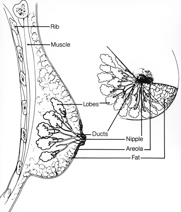 | 600 × 702 (74 KB) | S8600021 (talk | contribs) |
You cannot overwrite this file.
File usage
The following 11 pages use this file:
- 2009 Lecture 18
- 2010 Lecture 22
- ANAT2241 Female Reproductive System
- BGDA Practical - Female Reproductive Tract Histology
- BGDB Gastrointestinal - Activity 4
- BGDB Gastrointestinal - Postnatal
- Integumentary System - Mammary Gland Development
- M
- Normal Development - Milk
- REI - Reproductive Medicine Seminar 2018
- Royal Hospital for Women - Reproductive Medicine Seminar 2018