File:Liver-sinusiod cartoon.jpg
Liver-sinusiod_cartoon.jpg (600 × 523 pixels, file size: 51 KB, MIME type: image/jpeg)
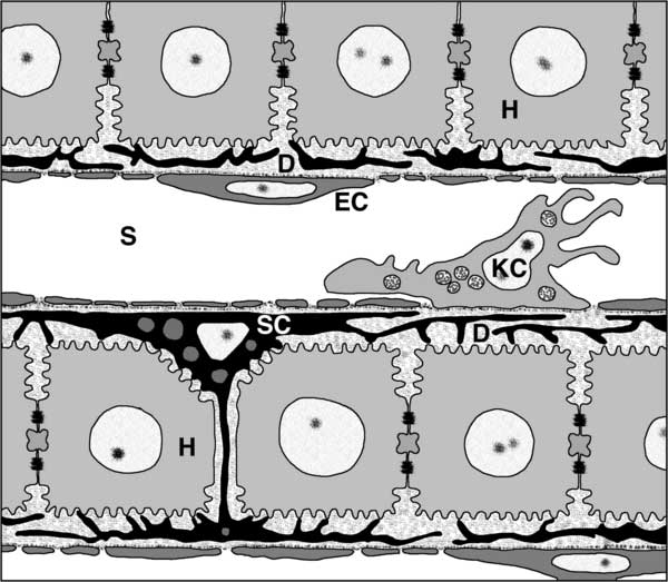
Architecture of the Liver Sinusoid
Liver sinusoids (S) are lined by fenestrated endothelia (EC) and interspersed Kupffer cells (KC), the resident macrophages of the liver. Stellate cells (SC), the major producers of liver ECM, are located inside the narrow space of Disse (D), which is formed by the sinusoidal cell layer and cords of hepatocytes (H).
Figure 1. Journal.pbio.0030192.g001.png
http://www.plosbiology.org/article/info%3Adoi%2F10.1371%2Fjournal.pbio.0030192
Citation: Frevert U, Engelmann S, Zougbédé S, Stange J, Ng B, et al. (2005) Intravital Observation of Plasmodium berghei Sporozoite Infection of the Liver. PLoS Biol 3(6): e192. doi:10.1371/journal.pbio.0030192
Academic Editor: Thomas Egwang, Medical Biotechnology Labs, Uganda
Received: December 7, 2004; Accepted: March 30, 2005; Published: May 24, 2005
Copyright: © 2005 Frevert et al. This is an open-access article distributed under the terms of the Creative Commons Attribution License, which permits unrestricted use, distribution, and reproduction in any medium, provided the original work is properly cited.
File history
Click on a date/time to view the file as it appeared at that time.
| Date/Time | Thumbnail | Dimensions | User | Comment | |
|---|---|---|---|---|---|
| current | 08:47, 1 May 2010 | | 600 × 523 (51 KB) | S8600021 (talk | contribs) | Architecture of the Liver Sinusoid Liver sinusoids (S) are lined by fenestrated endothelia (EC) and interspersed Kupffer cells (KC), the resident macrophages of the liver. Stellate cells (SC), the major producers of liver ECM, are located inside the narr |
You cannot overwrite this file.
File usage
There are no pages that use this file.