File:Lisser1911 fig30.jpg
From Embryology
Size of this preview: 643 × 600 pixels. Other resolution: 1,070 × 998 pixels.
Original file (1,070 × 998 pixels, file size: 249 KB, MIME type: image/jpeg)
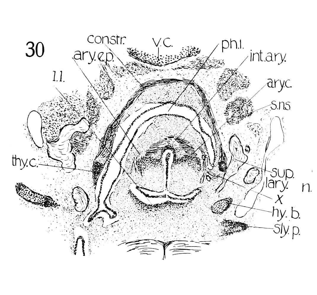
Fig. 30 Cross section of human Embryo 22 M. interarytaenoideus and aryepiglotticus
Embryo no. 22 (20 mm.) to show, especially, M. interarytaenoideus and aryepiglotticus. This section shows at the mark (x) the tendency to continuity between the laryngeal and pharj'ngeal musculature, as - mentioned by Strazza.
- Links: fig 1 | fig 2 | fig 3 | fig 4 | fig 5 | fig 6 | fig 7 | fig 8 | fig 9 | fig 10 | fig 11 | fig 12 | fig 13 | fig 14 | Lisser 1911
Reference
Lisser H. Studies on the development of the human larynx. (1911) Amer. J Anat. 12: 27-66.
Cite this page: Hill, M.A. (2024, April 27) Embryology Lisser1911 fig30.jpg. Retrieved from https://embryology.med.unsw.edu.au/embryology/index.php/File:Lisser1911_fig30.jpg
- © Dr Mark Hill 2024, UNSW Embryology ISBN: 978 0 7334 2609 4 - UNSW CRICOS Provider Code No. 00098G
File history
Click on a date/time to view the file as it appeared at that time.
| Date/Time | Thumbnail | Dimensions | User | Comment | |
|---|---|---|---|---|---|
| current | 20:54, 15 June 2016 | | 1,070 × 998 (249 KB) | Z8600021 (talk | contribs) |
You cannot overwrite this file.
File usage
The following 3 pages use this file: