File:Kollmann739.jpg

Original file (781 × 667 pixels, file size: 79 KB, MIME type: image/jpeg)
- This text is a Google translate computer generated translation and may contain many errors.
Images from - Atlas of the Development of Man (Volume 2)
(Handatlas der entwicklungsgeschichte des menschen)
- Kollmann Atlas 2: Gastrointestinal | Respiratory | Urogenital | Cardiovascular | Neural | Integumentary | Smell | Vision | Hearing | Kollmann Atlas 1 | Kollmann Atlas 2 | Julius Kollmann
- Links: Julius Kollman | Atlas Vol.1 | Atlas Vol.2 | Embryology History
| Historic Disclaimer - information about historic embryology pages |
|---|
 |
Reference
Kollmann JKE. Atlas of the Development of Man (Handatlas der entwicklungsgeschichte des menschen). (1907) Vol.1 and Vol. 2. Jena, Gustav Fischer. (1898).
Cite this page: Hill, M.A. (2024, May 15) Embryology Kollmann739.jpg. Retrieved from https://embryology.med.unsw.edu.au/embryology/index.php/File:Kollmann739.jpg
- © Dr Mark Hill 2024, UNSW Embryology ISBN: 978 0 7334 2609 4 - UNSW CRICOS Provider Code No. 00098G
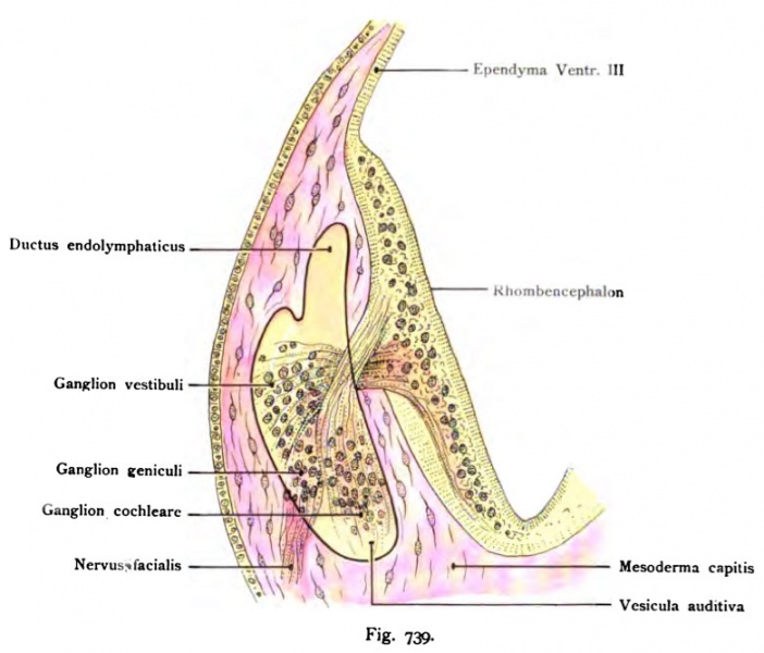
Fig. 739. Ursprung äes Nervus acusticus mit dem Ganglion vestibuläre und
dem Ganglion cochleare
später Ganglion spirale, bei dem menschlichen Embryo von 3 V» Wocheu ; dazu das Ganglion geniculi des Facialis und die Wurzel des nämlichen Nerven, alle drei
Ganglien zusammen benannt als Acustico-facialis-ganglion.
Die Lage der drei Ganglien ist durch Farben kenntlich gemacht. Das Hör- oder Labyrinthbläschen ist durch eine ausgezogene Linie angegeben. Die Ganglien des Acusticus hegen ihm an, der Facialisstamm wendet sich nach ab- wärts, um am Unterkiefer mit der Anlage des Platysma zusammenzutreffen. Als motorischer Nerv ist der Stamm des N. facialis rot tingiert.
File history
Click on a date/time to view the file as it appeared at that time.
| Date/Time | Thumbnail | Dimensions | User | Comment | |
|---|---|---|---|---|---|
| current | 12:29, 21 October 2011 | | 781 × 667 (79 KB) | S8600021 (talk | contribs) | {{Kollmann1907}} Category:Hearing Fig. 739. Ursprung äes Nervus acusticus mit dem Ganglion vestibuläre und dem Ganglion cochleare später Ganglion spirale, bei dem menschlichen Embryo von 3 V» Wocheu ; dazu das Ganglion geniculi des Faciali |
You cannot overwrite this file.
File usage
The following 2 pages use this file: