File:Kollmann725.jpg
Kollmann725.jpg (697 × 307 pixels, file size: 33 KB, MIME type: image/jpeg)
- This text is a Google translate computer generated translation and may contain many errors.
Images from - Atlas of the Development of Man (Volume 2)
(Handatlas der entwicklungsgeschichte des menschen)
- Kollmann Atlas 2: Gastrointestinal | Respiratory | Urogenital | Cardiovascular | Neural | Integumentary | Smell | Vision | Hearing | Kollmann Atlas 1 | Kollmann Atlas 2 | Julius Kollmann
- Links: Julius Kollman | Atlas Vol.1 | Atlas Vol.2 | Embryology History
| Historic Disclaimer - information about historic embryology pages |
|---|
 |
Reference
Kollmann JKE. Atlas of the Development of Man (Handatlas der entwicklungsgeschichte des menschen). (1907) Vol.1 and Vol. 2. Jena, Gustav Fischer. (1898).
Cite this page: Hill, M.A. (2024, April 27) Embryology Kollmann725.jpg. Retrieved from https://embryology.med.unsw.edu.au/embryology/index.php/File:Kollmann725.jpg
- © Dr Mark Hill 2024, UNSW Embryology ISBN: 978 0 7334 2609 4 - UNSW CRICOS Provider Code No. 00098G
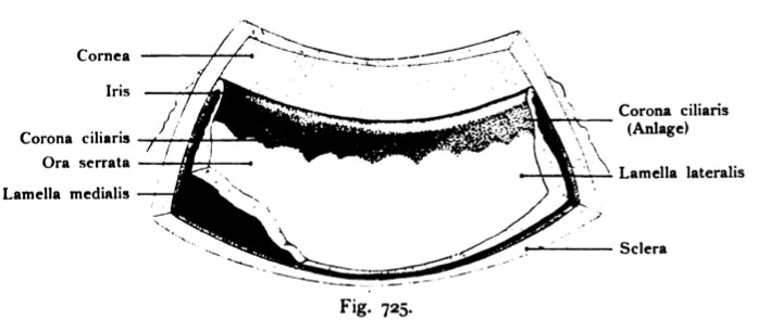
Fig. 725. Ora serrata mit Pars iridica retinae und vorderer Umschlassrand
der iooeren Lamelle der Retina
in die äußere Ljamelle bei einem menschlichen Fetus vom Ende des 3. Monates (4,8 cm Scheitelsteißlänge). Segment aus dem vorderen Umfang des Bulbus.
(Anatomische Sammlung in Basel)
Der Irisrand, in welchem später der Sphincter pupillae auftritt, erscheint wenig pigmentiert, weil die laterale Lamelle der Retina dort noch kein Pigment aufgenommen hat. Auf der dunklen folgenden Zone entwickeln sich die Pro- cessus ciliares, wie der Schnitt rechts an der Figur erkennen läßt. Dieser Teil der Pars ciliaris retinae erscheint stark pigmentiert. Der folgende Abschnitt, nicht minder pigmentiert, ist von der Ora serrata retinae überdeckt; an der unteren Ecke links ist die laterale Lamelle der Retina abgetragen, um die mediale sehen zu können. 40 mal vergr.
File history
Click on a date/time to view the file as it appeared at that time.
| Date/Time | Thumbnail | Dimensions | User | Comment | |
|---|---|---|---|---|---|
| current | 10:53, 21 October 2011 | | 697 × 307 (33 KB) | S8600021 (talk | contribs) | {{Kollmann1907}} Category:Vision Fig. 725. Ora serrata mit Pars iridica retinae und vorderer Umschlassrand der iooeren Lamelle der Retina in die äußere Ljamelle bei einem menschlichen Fetus vom Ende des 3. Monates (4,8 cm Scheitelsteißlänge |
You cannot overwrite this file.
File usage
The following page uses this file: