File:Kollmann719.jpg
Kollmann719.jpg (775 × 579 pixels, file size: 87 KB, MIME type: image/jpeg)
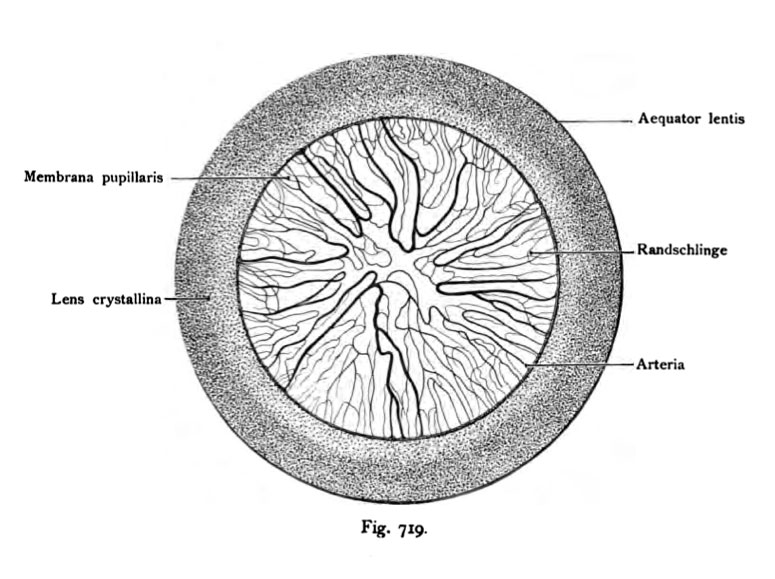
719. Membrane pupillaris
The iris is removed. From all over the area to push the vessel back to the center of the lens lateral pole which is itself free of the vessels is a sign of incipient recovery, which eliminates the normal state gradually the whole membrane. The arteries of the papillary membrane arise from the major circle of iris, the edge loops, the venous flow in the veins vorticosae.
- This text is a Google translate computer generated translation and may contain many errors.
Images from - Atlas of the Development of Man (Volume 2)
(Handatlas der entwicklungsgeschichte des menschen)
- Kollmann Atlas 2: Gastrointestinal | Respiratory | Urogenital | Cardiovascular | Neural | Integumentary | Smell | Vision | Hearing | Kollmann Atlas 1 | Kollmann Atlas 2 | Julius Kollmann
- Links: Julius Kollman | Atlas Vol.1 | Atlas Vol.2 | Embryology History
| Historic Disclaimer - information about historic embryology pages |
|---|
 |
Reference
Kollmann JKE. Atlas of the Development of Man (Handatlas der entwicklungsgeschichte des menschen). (1907) Vol.1 and Vol. 2. Jena, Gustav Fischer. (1898).
Cite this page: Hill, M.A. (2024, May 9) Embryology Kollmann719.jpg. Retrieved from https://embryology.med.unsw.edu.au/embryology/index.php/File:Kollmann719.jpg
- © Dr Mark Hill 2024, UNSW Embryology ISBN: 978 0 7334 2609 4 - UNSW CRICOS Provider Code No. 00098G
Fig. 719. Membrana pupUlaris
auf der vorderen Fläche der Linse aufliegend, injiziert; vom menschlichen Fetus
des 8. Monats.
(Nach O. Schultze.)
Die Iris ist entfernt. Von der ganzen Umgebung schieben sich die Gefäße gegen das Zentrum des lateralen Linsenpoles hin, der selbst von Gefäßen frei ist Das ist jedoch schon ein Zeichen beginnender Rückbildung, welche im normalen Zustand nach und nach die ganze Membran beseitigt. Die Arterien der Papillar- membran entspringen aus dem Circulus iridis major, die Randschlingen, die Venen fließen in die Venae vorticosae.
File history
Click on a date/time to view the file as it appeared at that time.
| Date/Time | Thumbnail | Dimensions | User | Comment | |
|---|---|---|---|---|---|
| current | 10:50, 21 October 2011 | | 775 × 579 (87 KB) | S8600021 (talk | contribs) | {{Kollmann1907}} Category:Vision Fig. 719. Membrana pupUlaris auf der vorderen Fläche der Linse aufliegend, injiziert; vom menschlichen Fetus des 8. Monats. (Nach O. Schultze.) Die Iris ist entfernt. Von der ganzen Umgebung schieben sich |
You cannot overwrite this file.
File usage
The following page uses this file: