File:Kollmann709.jpg
Kollmann709.jpg (781 × 592 pixels, file size: 132 KB, MIME type: image/jpeg)
- This text is a Google translate computer generated translation and may contain many errors.
Images from - Atlas of the Development of Man (Volume 2)
(Handatlas der entwicklungsgeschichte des menschen)
- Kollmann Atlas 2: Gastrointestinal | Respiratory | Urogenital | Cardiovascular | Neural | Integumentary | Smell | Vision | Hearing | Kollmann Atlas 1 | Kollmann Atlas 2 | Julius Kollmann
- Links: Julius Kollman | Atlas Vol.1 | Atlas Vol.2 | Embryology History
| Historic Disclaimer - information about historic embryology pages |
|---|
 |
Reference
Kollmann JKE. Atlas of the Development of Man (Handatlas der entwicklungsgeschichte des menschen). (1907) Vol.1 and Vol. 2. Jena, Gustav Fischer. (1898).
Cite this page: Hill, M.A. (2024, April 28) Embryology Kollmann709.jpg. Retrieved from https://embryology.med.unsw.edu.au/embryology/index.php/File:Kollmann709.jpg
- © Dr Mark Hill 2024, UNSW Embryology ISBN: 978 0 7334 2609 4 - UNSW CRICOS Provider Code No. 00098G
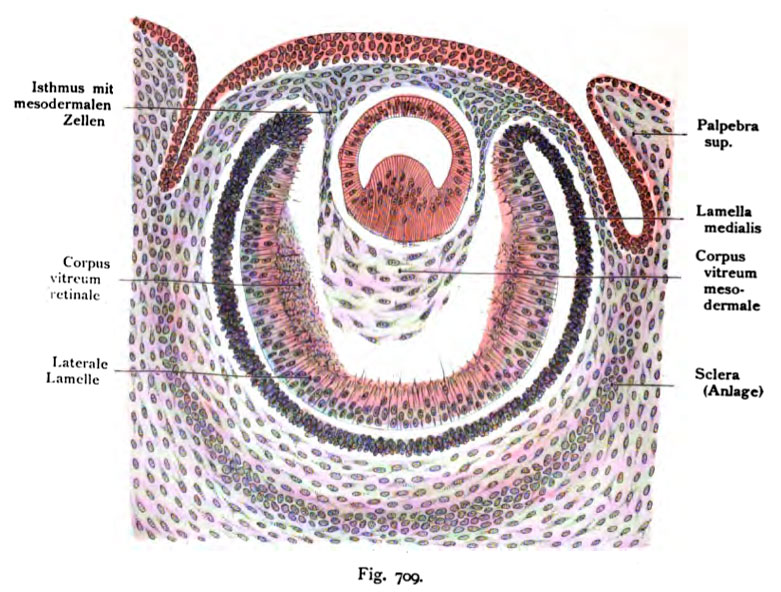
Fig. 709. Entstehung des Glaskörpers, Corpus vitreum, bei einem mensch- lichen Embryo von 15 mm Scheitelsteifliänge.
(Ende der 6. Woche.) (Anatomische Sammlung in Basel)
Die Konturen sind mit Hilfe des Projektionsapparates hergestellt. Die Schnittrichtung ist sagittal. Formol- Alkohol, Boraxkarmin, Balsam. Das Auge ist so orientiert, daß die Linse oben, das obere Lid rechts, das untere links liegt. Das Linsenbläschen ist von einem hellen Hof umgeben.' Zwischen Ektoderm und Linsenepithel haben sich Mesodermzellen vorgeschoben und zu einer kontinuierlichen Schicht verbunden, welche die erste Anlage der Tunica propria corneae darstellt und die Linse völlig von dem Ektoderm abdrängt. Der hintere Umfang der Linse ruht in dem mesodermalen Glaskörper, der oben am Isthmus mit dem Mesoderm der Tunica propria corneae zusammen- hängt und mit Bindesubstanzzellen ausgestattet ist. Der retinale Glaskörper ist längs der Seitenwand als feinstreifige Masse zu erkennen. Der leere Raum zwischen mesodermalem und retinalem Glaskörper ist wohl zum Teil auf Schrumpfung zurückzuführen, ebenso der helle Hof um das Linsenbläschen, wobei einige platte mesodermale Zellen auf dem Linsenbläschen sitzen blieben.
File history
Click on a date/time to view the file as it appeared at that time.
| Date/Time | Thumbnail | Dimensions | User | Comment | |
|---|---|---|---|---|---|
| current | 10:46, 21 October 2011 | | 781 × 592 (132 KB) | S8600021 (talk | contribs) | {{Kollmann1907}} Category:Vision Fig. 709. Entstehung des Glaskörpers, Corpus vitreum, bei einem mensch- lichen Embryo von 15 mm Scheitelsteifliänge. (Ende der 6. Woche.) (Anatomische Sammlung in Basel) Die Konturen sind mit Hilfe des Pr |
You cannot overwrite this file.
File usage
The following 2 pages use this file: