File:Kollmann698.jpg
Kollmann698.jpg (689 × 337 pixels, file size: 45 KB, MIME type: image/jpeg)
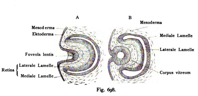
Fig. 698. The embryonic bulbus two human embryos at different stages of development of the eye cup
Both eyeballs from the 4th Weeks represented on average. The internal organization shows the following details: i vesicle optica secundaria äufeeren a double wall (lateral) and an inner (medial) lamella. Second A Linsengrübchen at the still open, according Koelliker. The rear wall of LinsengrObchens is also noticeable. Third The envelope by mesoderm. When B (van Bambecke) are the edges of the lens pit already grown and there is a lens vesicle is formed, which still related to the remaining ectoderm.
- This text is a Google translate computer generated translation and may contain many errors.
Images from - Atlas of the Development of Man (Volume 2)
(Handatlas der entwicklungsgeschichte des menschen)
- Kollmann Atlas 2: Gastrointestinal | Respiratory | Urogenital | Cardiovascular | Neural | Integumentary | Smell | Vision | Hearing | Kollmann Atlas 1 | Kollmann Atlas 2 | Julius Kollmann
- Links: Julius Kollman | Atlas Vol.1 | Atlas Vol.2 | Embryology History
| Historic Disclaimer - information about historic embryology pages |
|---|
 |
Reference
Kollmann JKE. Atlas of the Development of Man (Handatlas der entwicklungsgeschichte des menschen). (1907) Vol.1 and Vol. 2. Jena, Gustav Fischer. (1898).
Cite this page: Hill, M.A. (2024, April 27) Embryology Kollmann698.jpg. Retrieved from https://embryology.med.unsw.edu.au/embryology/index.php/File:Kollmann698.jpg
- © Dr Mark Hill 2024, UNSW Embryology ISBN: 978 0 7334 2609 4 - UNSW CRICOS Provider Code No. 00098G
Fig. 698. Der embryonale Bulbus zweier menschlichen Embryonen auf verschiedenen Entwicklungsstufen des Augenbechers.
Beide Bulbi aus der 4. Woche, im Durchschnitt dargestellt. Die innere Organisation zeigt folgende Einzelheiten: i. Die Vesicula optica secundaria mit doppelter Wandung einer äufeeren (lateralen) und einer inneren (medialen) Lamelle. 2. Bei A das noch offene Linsengrübchen, nach Koelliker. Die hintere Wand des LinsengrObchens ist ebenfalls bemerkbar. 3. Die Umhüllung durch Mesoderm. Bei B (nach van Bambecke) sind die Ränder des Linsen- grübchens bereits verwachsen und es ist ein Linsenbläschen entstanden, das aber noch mit dem übrigen Ektoderm zusammenhängt.
- Note - This image was originally uploaded as part of an undergraduate science student project and may contain inaccuracies in either description or acknowledgements. Students have been advised in writing concerning the reuse of content and may accidentally have misunderstood the original terms of use. If image reuse on this non-commercial educational site infringes your existing copyright, please contact the site editor for immediate removal.
File history
Click on a date/time to view the file as it appeared at that time.
| Date/Time | Thumbnail | Dimensions | User | Comment | |
|---|---|---|---|---|---|
| current | 10:40, 21 October 2011 | | 689 × 337 (45 KB) | S8600021 (talk | contribs) | {{Kollmann1907}} Category:Vision FiS. 698. Der embryonale Bulbus zweier menschlichen Embryonen auf ver- schiedenen Entwicklungsstufen des Augenbechers. Beide Bulbi aus der 4. Woche, im Durchschnitt dargestellt. Die innere Organisation zeigt f |
You cannot overwrite this file.
File usage
The following 3 pages use this file: