File:Kollmann695.jpg
Kollmann695.jpg (734 × 519 pixels, file size: 45 KB, MIME type: image/jpeg)
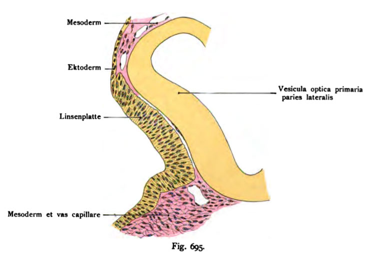
Figure 695 The lens unit with a rabbit embryo
(According to Rabl.)
The strongly bulging lateral wall of the primary optic vesicle is covered by a fairly well-defined lens plate, a direct continuation of the ectoderm. The lens plate ventrally slightly recessed and is a is high, narrow columnar cells. Between optic vesicle and the lens pit are some flattened spindle-shaped cells. In the adjoining mesoderm are cross-sections of capillaries. In the whole extent of the lateral surface considered, the lens plate has a recess. It therefore describes it as an Linsengrübchen (foveola lentis).
- This text is a Google translate computer generated translation and may contain many errors.
Images from - Atlas of the Development of Man (Volume 2)
(Handatlas der entwicklungsgeschichte des menschen)
- Kollmann Atlas 2: Gastrointestinal | Respiratory | Urogenital | Cardiovascular | Neural | Integumentary | Smell | Vision | Hearing | Kollmann Atlas 1 | Kollmann Atlas 2 | Julius Kollmann
- Links: Julius Kollman | Atlas Vol.1 | Atlas Vol.2 | Embryology History
| Historic Disclaimer - information about historic embryology pages |
|---|
 |
Reference
Kollmann JKE. Atlas of the Development of Man (Handatlas der entwicklungsgeschichte des menschen). (1907) Vol.1 and Vol. 2. Jena, Gustav Fischer. (1898).
Cite this page: Hill, M.A. (2024, May 11) Embryology Kollmann695.jpg. Retrieved from https://embryology.med.unsw.edu.au/embryology/index.php/File:Kollmann695.jpg
- © Dr Mark Hill 2024, UNSW Embryology ISBN: 978 0 7334 2609 4 - UNSW CRICOS Provider Code No. 00098G
Fig. 695. Die Linsenanlage bei einem lOtägisen Kaninchenembryo.
(Nach Rabl.)
Die stark vorgewölbte laterale Wand der primären Augenblase ist von einer ziemlich gut abgegrenzten Linsenplatte bedeckt, eine direkte Fortsetzung des Ektoderms. Die Linsenplatte ist ventralwärts etwas vertieft und besteht a|is hohen schmalen Zylinderzellen. Zwischen Augenblase und der Linsengrube liegen einige plattgedrückte spindelförmige Zellen. In dem anstoßenden Meso- derm befinden sich Querschnitte von Kapillaren. In dem ganzen Umfang von der lateralen Fläche betrachtet, besitzt die Linsenplatte eine Vertiefung. Man spricht deshalb schon von einem Linsengrübchen (Foveola lentis).
File history
Click on a date/time to view the file as it appeared at that time.
| Date/Time | Thumbnail | Dimensions | User | Comment | |
|---|---|---|---|---|---|
| current | 10:38, 21 October 2011 | | 734 × 519 (45 KB) | S8600021 (talk | contribs) | {{Kollmann1907}} Category:Vision Fig. 695. Die Linsenanlage bei einem lOtägisen Kaninchenembryo. (Nach Rabl.) Die stark vorgewölbte laterale Wand der primären Augenblase ist von einer ziemlich gut abgegrenzten Linsenplatte bedeckt, eine dir |
You cannot overwrite this file.
File usage
The following 3 pages use this file: