File:Kollmann607.jpg
Kollmann607.jpg (706 × 560 pixels, file size: 85 KB, MIME type: image/jpeg)
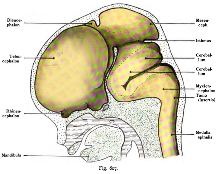
Fig. 607. Neural tube of a human embryo of 22 mm CRL about 7 weeks old
(Anatomical Collection in Basel)
The vesicle of the telencephalon has now grown considerably and covers almost the entire dorsal diencephalon. The midbrain starts at this upright position of the embryonic head and is still the highest point of the crown. Then follows the hindbrain and hindbrain associated with bend of the bridge together. This curvature is located at the mid-brain and would almost touch it, if the middle cranial beams between them were not present. In later stages, this has shortened and is more basal withdrawn. The space of the telencephalon is later to form the lateral ventricle, the cavity of the diencephalon to the ventriculus tertius, and the channel to the midbrain aqueduct. The space of the metencephalon forms the fourth ventricle and the floor in the space at the back of the myelencephalon section on the fourth ventricle.
- This text is a Google translate computer generated translation and may contain many errors.
Images from - Atlas of the Development of Man (Volume 2)
(Handatlas der entwicklungsgeschichte des menschen)
- Kollmann Atlas 2: Gastrointestinal | Respiratory | Urogenital | Cardiovascular | Neural | Integumentary | Smell | Vision | Hearing | Kollmann Atlas 1 | Kollmann Atlas 2 | Julius Kollmann
- Links: Julius Kollman | Atlas Vol.1 | Atlas Vol.2 | Embryology History
| Historic Disclaimer - information about historic embryology pages |
|---|
 |
Reference
Kollmann JKE. Atlas of the Development of Man (Handatlas der entwicklungsgeschichte des menschen). (1907) Vol.1 and Vol. 2. Jena, Gustav Fischer. (1898).
Cite this page: Hill, M.A. (2024, April 28) Embryology Kollmann607.jpg. Retrieved from https://embryology.med.unsw.edu.au/embryology/index.php/File:Kollmann607.jpg
- © Dr Mark Hill 2024, UNSW Embryology ISBN: 978 0 7334 2609 4 - UNSW CRICOS Provider Code No. 00098G
Fig. 607. Himrohr eines menschlichen Embryo von 22 mm Nackenlänge,
etwa 7 Wochen alt. (Anatomische Sammlung in Basel)
Das Großhimbläschen des Telencephalon ist jetzt beträchtlich gewachsen und bedeckt dorsal nahezu das ganze Zwischenhirn. Das Mittelhirn nimmt bei dieser aufrechten Stellung des embryonalen Kopfes noch immer den höchsten Punkt des Scheitels ein. Dann folgt Hinterhirn und Nachhirn, die durch die Brückenkrümmung miteinander zusammenhängen. Diese Krümmung liegt dem Zwischenhirn nahezu an und würde es berühren, wenn nicht der mittlere Schädelbalken sich dazwischen befände. In späteren Stadien hat er sich verkürzt und mehr basalwärts zurückgezogen. Die Höhle des Telencephalon wird später zu dem Ventriculus lateralis, die Höhle des Diencephalon zum Ventriculus tertius; der Kanal im Mesencephalon zum Aquaeductus, die Höhle des Metencephalon zum Ventriculus quartus und der Boden in der Höhle des Myelencephalon im hinteren Abschnitt zur Rautengrube.
File history
Click on a date/time to view the file as it appeared at that time.
| Date/Time | Thumbnail | Dimensions | User | Comment | |
|---|---|---|---|---|---|
| current | 16:54, 17 October 2011 | | 706 × 560 (85 KB) | S8600021 (talk | contribs) | {{Kollmann1907}} |
You cannot overwrite this file.
File usage
The following 2 pages use this file: